A convenient, reliable, and fast acoustic pressure field measurement method for magnetic resonance-guided high-intensity focused ultrasound systems with phased array transducers.
Satya V V N Kothapalli, Ari Partanen, Lifei Zhu, Michael B Altman, H Michael Gach, Dennis E Hallahan, Hong Chen
{"title":"A convenient, reliable, and fast acoustic pressure field measurement method for magnetic resonance-guided high-intensity focused ultrasound systems with phased array transducers.","authors":"Satya V V N Kothapalli, Ari Partanen, Lifei Zhu, Michael B Altman, H Michael Gach, Dennis E Hallahan, Hong Chen","doi":"10.1186/s40349-018-0113-7","DOIUrl":null,"url":null,"abstract":"<p><strong>Background: </strong>With the expanding applications of magnetic resonance-guided high-intensity focused ultrasound (MR-HIFU), there is an urgent need for a convenient, reliable, and fast acoustic pressure field measurement method to aid treatment protocol design, ensure consistent and safe operation of the transducer, and facilitate regulatory approval of new techniques. Herein, we report a method for acoustic pressure field characterization of MR-HIFU systems with multi-element phased array transducers. This method integrates fiber-optic hydrophone measurements and electronic steering of the ultrasound beam with MRI-assisted HIFU focus alignment to the fiber tip.</p><p><strong>Methods: </strong>A clinical MR-HIFU system (Sonalleve V2, Profound Medical Inc., Mississauga, Canada) was used to assess the proposed method. A fiber-optic hydrophone was submerged in a degassed water bath, and the fiber tip location was traced using MRI. Subsequently, the nominal transducer focal point indicated on the MR-HIFU therapy planning software was positioned at the fiber tip, and the HIFU focus was electronically steered around the fiber tip within a 3D volume for 3D pressure field mapping, eliminating the need for an additional, expensive, and MRI-compatible 3D positioning stage. The peak positive and negative pressures were measured at the focus and validated using a standard hydrophone measurement setup outside the MRI magnet room.</p><p><strong>Results: </strong>We found that the initial MRI-assisted HIFU focus alignment had an average offset of 2.23 ± 1.33 mm from the fiber tip as identified by the 3D pressure field mapping. MRI guidance and electronic beam steering allowed 3D focus localization within ~ 1 h, i.e., faster than the typical time required using the standard laboratory setup (~ 3-4 h). Acoustic pressures measured using the proposed method were not significantly different from those obtained with the standard laboratory hydrophone measurements.</p><p><strong>Conclusions: </strong>In conclusion, our method offers a convenient, reliable, and fast acoustic pressure field characterization tool for MR-HIFU systems with phased array transducers.</p>","PeriodicalId":90245,"journal":{"name":"Journal of therapeutic ultrasound","volume":"6 ","pages":"5"},"PeriodicalIF":0.0000,"publicationDate":"2018-07-02","publicationTypes":"Journal Article","fieldsOfStudy":null,"isOpenAccess":false,"openAccessPdf":"https://www.ncbi.nlm.nih.gov/pmc/articles/PMC6027582/pdf/","citationCount":"0","resultStr":null,"platform":"Semanticscholar","paperid":null,"PeriodicalName":"Journal of therapeutic ultrasound","FirstCategoryId":"1085","ListUrlMain":"https://doi.org/10.1186/s40349-018-0113-7","RegionNum":0,"RegionCategory":null,"ArticlePicture":[],"TitleCN":null,"AbstractTextCN":null,"PMCID":null,"EPubDate":"2018/1/1 0:00:00","PubModel":"eCollection","JCR":"","JCRName":"","Score":null,"Total":0}
引用次数: 0
Abstract
Background: With the expanding applications of magnetic resonance-guided high-intensity focused ultrasound (MR-HIFU), there is an urgent need for a convenient, reliable, and fast acoustic pressure field measurement method to aid treatment protocol design, ensure consistent and safe operation of the transducer, and facilitate regulatory approval of new techniques. Herein, we report a method for acoustic pressure field characterization of MR-HIFU systems with multi-element phased array transducers. This method integrates fiber-optic hydrophone measurements and electronic steering of the ultrasound beam with MRI-assisted HIFU focus alignment to the fiber tip.
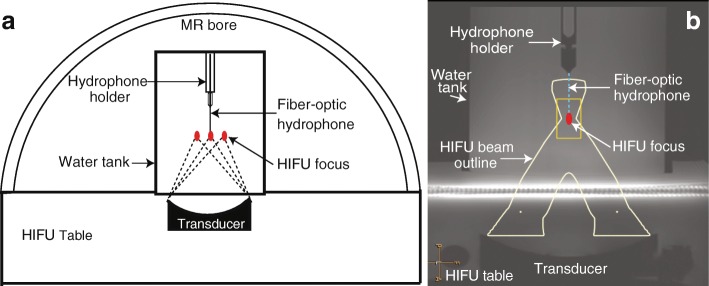
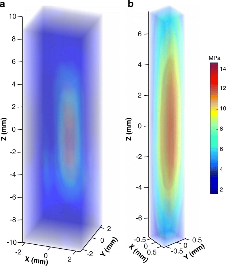
Methods: A clinical MR-HIFU system (Sonalleve V2, Profound Medical Inc., Mississauga, Canada) was used to assess the proposed method. A fiber-optic hydrophone was submerged in a degassed water bath, and the fiber tip location was traced using MRI. Subsequently, the nominal transducer focal point indicated on the MR-HIFU therapy planning software was positioned at the fiber tip, and the HIFU focus was electronically steered around the fiber tip within a 3D volume for 3D pressure field mapping, eliminating the need for an additional, expensive, and MRI-compatible 3D positioning stage. The peak positive and negative pressures were measured at the focus and validated using a standard hydrophone measurement setup outside the MRI magnet room.
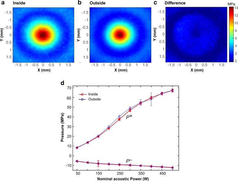
Results: We found that the initial MRI-assisted HIFU focus alignment had an average offset of 2.23 ± 1.33 mm from the fiber tip as identified by the 3D pressure field mapping. MRI guidance and electronic beam steering allowed 3D focus localization within ~ 1 h, i.e., faster than the typical time required using the standard laboratory setup (~ 3-4 h). Acoustic pressures measured using the proposed method were not significantly different from those obtained with the standard laboratory hydrophone measurements.
Conclusions: In conclusion, our method offers a convenient, reliable, and fast acoustic pressure field characterization tool for MR-HIFU systems with phased array transducers.

 求助内容:
求助内容: 应助结果提醒方式:


