Fereydoon Sargolzaei Aval, Mohammad Reza Arab, Narjes Sargolzaei, Fateme Noushadi, Abdolsamad Eteghadi, Asadollah Keykhaei, Foroug Sargolzaei Aval, Azim Hedayat Pour
{"title":"Efficacy of Octacalcium Phosphate and Octacalcium Phosphate/Gelatin Composite on the Repair of Critical-Sized Calvarial Defects in Rats.","authors":"Fereydoon Sargolzaei Aval, Mohammad Reza Arab, Narjes Sargolzaei, Fateme Noushadi, Abdolsamad Eteghadi, Asadollah Keykhaei, Foroug Sargolzaei Aval, Azim Hedayat Pour","doi":"","DOIUrl":null,"url":null,"abstract":"<p><strong>Objectives: </strong>The healing of bone defects in the craniofacial region is an important clinical issue. We aimed to compare the effects of octacalcium phosphate (OCP) and the combination of OCP/gelatin (OCP/Gel) on calvarial bone regeneration in rats.</p><p><strong>Materials and methods: </strong>In this study, 72 male Sprague Dawley rats were randomly assigned to the OCP (n=24), OCP/Gel (n=24), and control groups (n=24). Lesions with a diameter of 9 mm were created in the parietal bone and were filled with 9-mg OCP and OCP/Gel disks. In the control group, no substance was implanted in the defect. Sampling was performed on days 10, 14, 21, and 28 after the implantation. After tissue processing, 5-μm sections were prepared and stained by hematoxylin and eosin (H&E) stain. The sections were studied, and the volume fraction of the newly formed bone was assessed by Kruskal-Wallis test at a significance level of 0.05.</p><p><strong>Results: </strong>In the experimental groups, new bone formation was detected at the margins of the defects 10 days after the implantation. With the progression of the healing process, the newly formed bone covered greater areas of the defects and developed a more mature structure. In the control group, the defects were primarily filled with a dense connective tissue with small islands of new bone. The results of histomorphometric assessments showed that the volume of the newly formed bone in the experimental groups had a significant statistical difference with that in the control group (P<0.001).</p><p><strong>Conclusions: </strong>The OCP/Gel composite can be useful in the healing process of calvarial bone defects.</p>","PeriodicalId":30286,"journal":{"name":"Journal of Dentistry of Tehran University of Medical Sciences","volume":"15 2","pages":"86-96"},"PeriodicalIF":0.0000,"publicationDate":"2018-03-01","publicationTypes":"Journal Article","fieldsOfStudy":null,"isOpenAccess":false,"openAccessPdf":"https://www.ncbi.nlm.nih.gov/pmc/articles/PMC6026312/pdf/","citationCount":"0","resultStr":null,"platform":"Semanticscholar","paperid":null,"PeriodicalName":"Journal of Dentistry of Tehran University of Medical Sciences","FirstCategoryId":"1085","ListUrlMain":"","RegionNum":0,"RegionCategory":null,"ArticlePicture":[],"TitleCN":null,"AbstractTextCN":null,"PMCID":null,"EPubDate":"","PubModel":"","JCR":"","JCRName":"","Score":null,"Total":0}
引用次数: 0
Abstract
Objectives: The healing of bone defects in the craniofacial region is an important clinical issue. We aimed to compare the effects of octacalcium phosphate (OCP) and the combination of OCP/gelatin (OCP/Gel) on calvarial bone regeneration in rats.
Materials and methods: In this study, 72 male Sprague Dawley rats were randomly assigned to the OCP (n=24), OCP/Gel (n=24), and control groups (n=24). Lesions with a diameter of 9 mm were created in the parietal bone and were filled with 9-mg OCP and OCP/Gel disks. In the control group, no substance was implanted in the defect. Sampling was performed on days 10, 14, 21, and 28 after the implantation. After tissue processing, 5-μm sections were prepared and stained by hematoxylin and eosin (H&E) stain. The sections were studied, and the volume fraction of the newly formed bone was assessed by Kruskal-Wallis test at a significance level of 0.05.
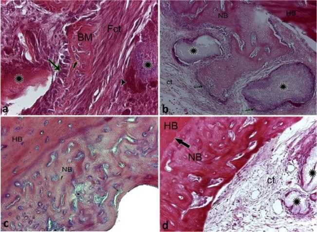
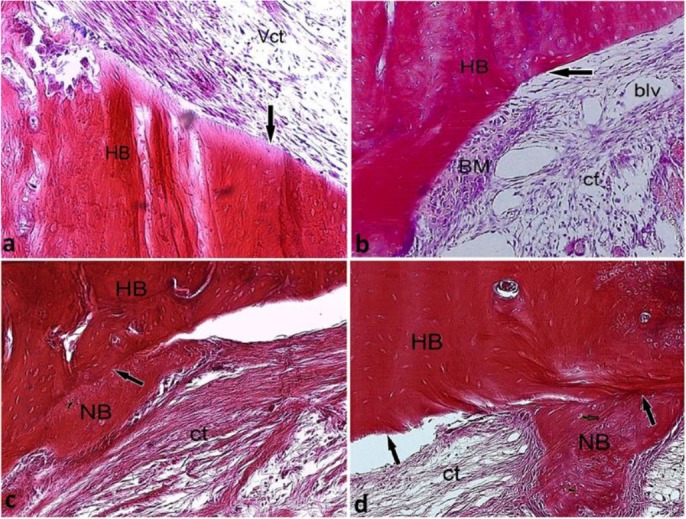
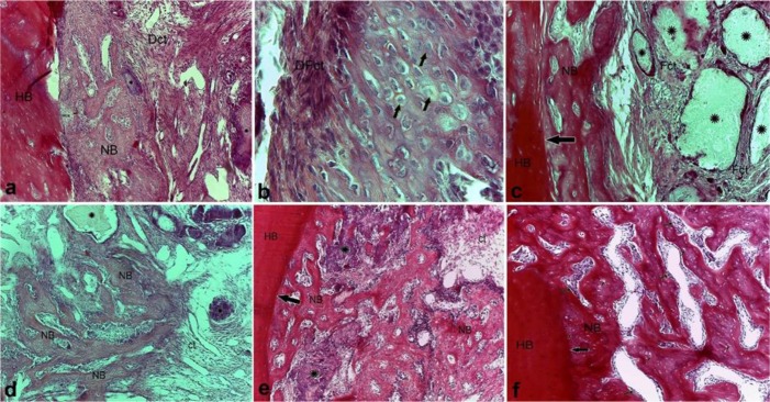
Results: In the experimental groups, new bone formation was detected at the margins of the defects 10 days after the implantation. With the progression of the healing process, the newly formed bone covered greater areas of the defects and developed a more mature structure. In the control group, the defects were primarily filled with a dense connective tissue with small islands of new bone. The results of histomorphometric assessments showed that the volume of the newly formed bone in the experimental groups had a significant statistical difference with that in the control group (P<0.001).
Conclusions: The OCP/Gel composite can be useful in the healing process of calvarial bone defects.

 求助内容:
求助内容: 应助结果提醒方式:


