{"title":"Adherence of <i>Streptococcus Mutans</i> to Microhybrid and Nanohybrid Resin Composites and Dental Amalgam: An In Vitro Study.","authors":"Fariba Motevasselian, Ensieh Zibafar, Esmail Yassini, Mansoreh Mirzaei, Naser Pourmirhoseni","doi":"","DOIUrl":null,"url":null,"abstract":"<p><strong>Objectives: </strong><i>Streptococcus mutans</i> (<i>S. mutans</i>) is a cariogenic microorganism. The restorative materials which harbor a biofilm with high levels of <i>S. mutans</i> can accelerate the occurrence of dental caries. The purpose of this study was to evaluate the influence of different restorative materials on <i>S. mutans</i> colonization in a simple in-vitro biofilm formation model.</p><p><strong>Materials and methods: </strong>Thirteen discs of each material (nanohybrid resin composite, microhybrid resin composite, and amalgam) were prepared, polished, and sterilized in a gamma radiation chamber. The saliva-free specimens were exposed to the <i>S. mutans</i> bacterial suspension (0.5 McFarland) and were incubated for 4 hours. Afterwards, the specimens were rinsed and sonicated in normal saline. 10μl of the obtained suspension was cultured in a sterile blood agar medium. After 24 hours, the number of colony forming units (CFU) of <i>S. mutans</i> was counted. A sterility test control was considered for each group of materials. The data were analyzed by one-way ANOVA at 5% significance level.</p><p><strong>Results: </strong>The means and standard deviations of the logarithmic values of the colonies on the surfaces of amalgam, microhybrid, and nanohybrid resin composites were equal to 3.76±0.64, 3.91±0.52 and 3.34±0.74, respectively.</p><p><strong>Conclusions: </strong>There were no significant differences between the restorative materials in terms of <i>S. mutans</i> adhesion rate. The evaluated resin composites showed comparable numbers of CFUs, which could imply the importance of the polishing procedures.</p>","PeriodicalId":30286,"journal":{"name":"Journal of Dentistry of Tehran University of Medical Sciences","volume":"14 6","pages":"337-343"},"PeriodicalIF":0.0000,"publicationDate":"2017-11-01","publicationTypes":"Journal Article","fieldsOfStudy":null,"isOpenAccess":false,"openAccessPdf":"https://www.ncbi.nlm.nih.gov/pmc/articles/PMC6015592/pdf/","citationCount":"0","resultStr":null,"platform":"Semanticscholar","paperid":null,"PeriodicalName":"Journal of Dentistry of Tehran University of Medical Sciences","FirstCategoryId":"1085","ListUrlMain":"","RegionNum":0,"RegionCategory":null,"ArticlePicture":[],"TitleCN":null,"AbstractTextCN":null,"PMCID":null,"EPubDate":"","PubModel":"","JCR":"","JCRName":"","Score":null,"Total":0}
引用次数: 0
Abstract
Objectives: Streptococcus mutans (S. mutans) is a cariogenic microorganism. The restorative materials which harbor a biofilm with high levels of S. mutans can accelerate the occurrence of dental caries. The purpose of this study was to evaluate the influence of different restorative materials on S. mutans colonization in a simple in-vitro biofilm formation model.
Materials and methods: Thirteen discs of each material (nanohybrid resin composite, microhybrid resin composite, and amalgam) were prepared, polished, and sterilized in a gamma radiation chamber. The saliva-free specimens were exposed to the S. mutans bacterial suspension (0.5 McFarland) and were incubated for 4 hours. Afterwards, the specimens were rinsed and sonicated in normal saline. 10μl of the obtained suspension was cultured in a sterile blood agar medium. After 24 hours, the number of colony forming units (CFU) of S. mutans was counted. A sterility test control was considered for each group of materials. The data were analyzed by one-way ANOVA at 5% significance level.
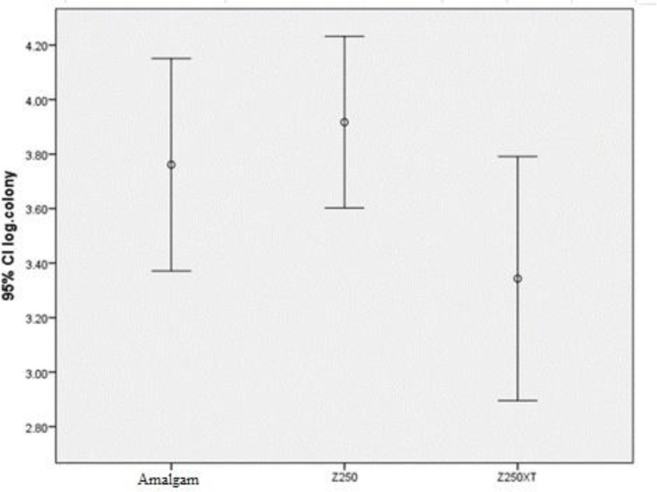
Results: The means and standard deviations of the logarithmic values of the colonies on the surfaces of amalgam, microhybrid, and nanohybrid resin composites were equal to 3.76±0.64, 3.91±0.52 and 3.34±0.74, respectively.
Conclusions: There were no significant differences between the restorative materials in terms of S. mutans adhesion rate. The evaluated resin composites showed comparable numbers of CFUs, which could imply the importance of the polishing procedures.

 求助内容:
求助内容: 应助结果提醒方式:


