Alberto Naoki Miyazaki, Luciana Andrade Silva, Pedro Doneux Santos, Guilherme do Val Sella, Leonardo Hideto Nagaya, Sergio Luiz Checchia
{"title":"Hill–Sachs lesion measurement with tridimensional models in anterior shoulder instability","authors":"Alberto Naoki Miyazaki, Luciana Andrade Silva, Pedro Doneux Santos, Guilherme do Val Sella, Leonardo Hideto Nagaya, Sergio Luiz Checchia","doi":"10.1016/j.rboe.2018.03.008","DOIUrl":null,"url":null,"abstract":"<div><h3>Objective</h3><p>To evaluate the reproducibility and repeatability of Hill–Sachs lesion measurement from computed tomography images, with computer software and tridimensional prototype.</p></div><div><h3>Methods</h3><p>Three-dimensional models were made from computed tomography images from 14 patients with anterior shoulder instability, using InVesalius 3.0® software. Hill–Sachs lesions were measured with Rhinocerus 5.0® software with pre-determined position. Mid-lateral distance, perpendicular to humeral shaft, cranial-caudal distance, parallel to humeral shaft, and the longitudinal distance of the lesion were measured. Using the Printer-ZP 310 three-dimensional printer, plaster models were made. To measure the Hill–Sachs lesion, a calibrated universal digital caliper was used in the same way as the software.</p></div><div><h3>Results</h3><p>There was intra-observer and inter-observer variability for measurement of the same model. Observers did not perform the measurements in a similar way, showing difficulty to use the method (<em>p</em> <!--><<!--> <!-->0.05). Using the software to measure the mid-lateral distance, as well as in the measurement with the caliper, the model type influenced the measurements for each of the observers, rendering the method invalid (<em>p</em> <!--><<!--> <!-->0.05).</p></div><div><h3>Conclusion</h3><p>There was no reproducibility and repeatability for Hill–Sachs lesion measurement between plaster models and software models.</p></div>","PeriodicalId":101095,"journal":{"name":"Revista Brasileira de Ortopedia (English Edition)","volume":"53 3","pages":"Pages 357-363"},"PeriodicalIF":0.0000,"publicationDate":"2018-05-01","publicationTypes":"Journal Article","fieldsOfStudy":null,"isOpenAccess":false,"openAccessPdf":"https://sci-hub-pdf.com/10.1016/j.rboe.2018.03.008","citationCount":"3","resultStr":null,"platform":"Semanticscholar","paperid":null,"PeriodicalName":"Revista Brasileira de Ortopedia (English Edition)","FirstCategoryId":"1085","ListUrlMain":"https://www.sciencedirect.com/science/article/pii/S2255497118300478","RegionNum":0,"RegionCategory":null,"ArticlePicture":[],"TitleCN":null,"AbstractTextCN":null,"PMCID":null,"EPubDate":"","PubModel":"","JCR":"","JCRName":"","Score":null,"Total":0}
引用次数: 3
Abstract
Objective
To evaluate the reproducibility and repeatability of Hill–Sachs lesion measurement from computed tomography images, with computer software and tridimensional prototype.
Methods
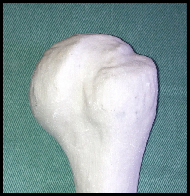
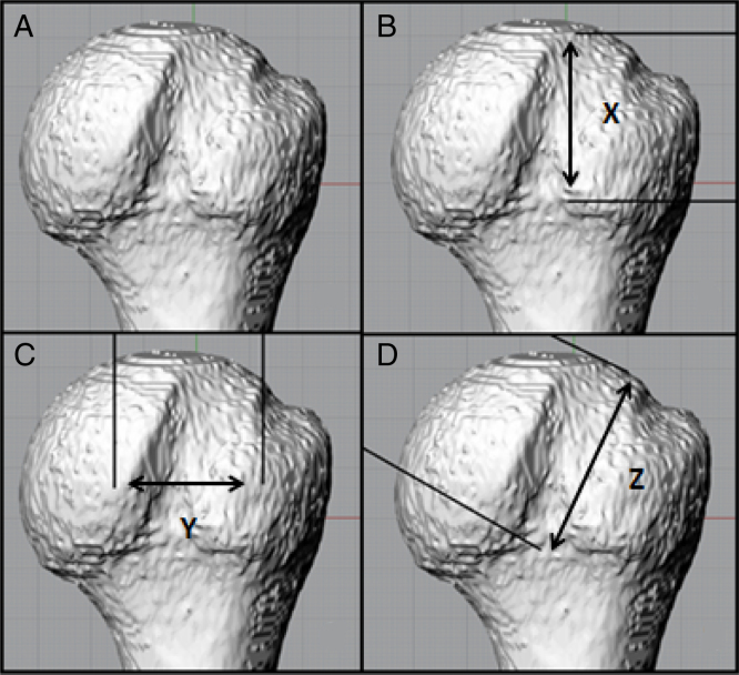
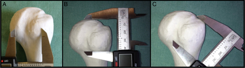
Three-dimensional models were made from computed tomography images from 14 patients with anterior shoulder instability, using InVesalius 3.0® software. Hill–Sachs lesions were measured with Rhinocerus 5.0® software with pre-determined position. Mid-lateral distance, perpendicular to humeral shaft, cranial-caudal distance, parallel to humeral shaft, and the longitudinal distance of the lesion were measured. Using the Printer-ZP 310 three-dimensional printer, plaster models were made. To measure the Hill–Sachs lesion, a calibrated universal digital caliper was used in the same way as the software.
Results
There was intra-observer and inter-observer variability for measurement of the same model. Observers did not perform the measurements in a similar way, showing difficulty to use the method (p < 0.05). Using the software to measure the mid-lateral distance, as well as in the measurement with the caliper, the model type influenced the measurements for each of the observers, rendering the method invalid (p < 0.05).
Conclusion
There was no reproducibility and repeatability for Hill–Sachs lesion measurement between plaster models and software models.

 求助内容:
求助内容: 应助结果提醒方式:


