{"title":"Effect of Filtration and Thickness of Cross-Sections of Cone Beam Computed Tomography Images on Detection of Proximal Caries.","authors":"Mehrdad Abdinian, Rahman Nazeri, Marzieh Ghaiour","doi":"","DOIUrl":null,"url":null,"abstract":"<p><strong>Objectives: </strong>When a patient has cone beam computed tomography (CBCT) images based on the treatment plan, it is possible to use these images for evaluation of caries, and there is no need for new radiographs, according to the \"as low as reasonably achievable\" (ALARA) principle. The aim of this study was to determine the effect of filtration and thickness of CBCT cross-sections on detection of proximal caries.</p><p><strong>Materials and methods: </strong>In this in-vitro study, 100 teeth were placed in the dental sockets of a dry skull, and were fixed in normal proximal contacts. CBCT images were taken and were evaluated by two observers on the panoramic view at 1-, 3- and 5-mm-thick cross-sections, with the use of filtrations 0, 1 and 2. Afterwards, the samples were sectioned and underwent a histological evaluation. McNemar's test was used to compare the findings on CBCT images and histological evaluation. Receiver operating characteristic (ROC) curves and logistic regression were used to evaluate the diagnostic accuracy of different cross-sections.</p><p><strong>Results: </strong>The maximum AZ-value was achieved at 3-mm thickness/filtration 2. However, the differences between 1-mm thickness/filtration 2 and 1-mm thickness/filtration 1 were not significant (P=0.728 and 0.868, respectively). The minimum AZ-value was achieved at 5-mm thickness/filtration 0.</p><p><strong>Conclusions: </strong>Although CBCT is not sufficiently effective in detecting caries, the best cross-sections for detection of proximal caries were achieved at 3-mm thickness/filtration 2, 1-mm thickness/filtration 2 and 1-mm thickness/filtration 1.</p>","PeriodicalId":30286,"journal":{"name":"Journal of Dentistry of Tehran University of Medical Sciences","volume":"14 4","pages":"223-230"},"PeriodicalIF":0.0000,"publicationDate":"2017-07-01","publicationTypes":"Journal Article","fieldsOfStudy":null,"isOpenAccess":false,"openAccessPdf":"https://www.ncbi.nlm.nih.gov/pmc/articles/PMC5745226/pdf/","citationCount":"0","resultStr":null,"platform":"Semanticscholar","paperid":null,"PeriodicalName":"Journal of Dentistry of Tehran University of Medical Sciences","FirstCategoryId":"1085","ListUrlMain":"","RegionNum":0,"RegionCategory":null,"ArticlePicture":[],"TitleCN":null,"AbstractTextCN":null,"PMCID":null,"EPubDate":"","PubModel":"","JCR":"","JCRName":"","Score":null,"Total":0}
引用次数: 0
Abstract
Objectives: When a patient has cone beam computed tomography (CBCT) images based on the treatment plan, it is possible to use these images for evaluation of caries, and there is no need for new radiographs, according to the "as low as reasonably achievable" (ALARA) principle. The aim of this study was to determine the effect of filtration and thickness of CBCT cross-sections on detection of proximal caries.
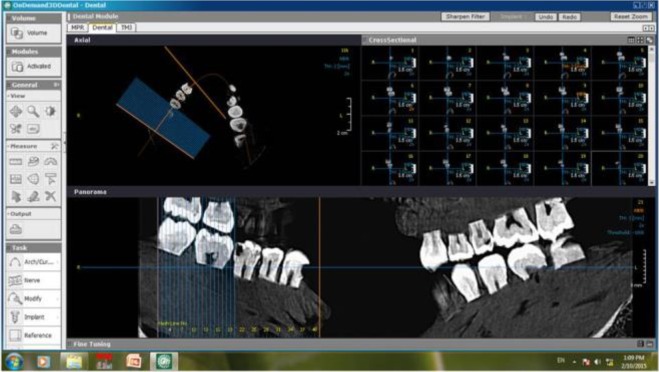
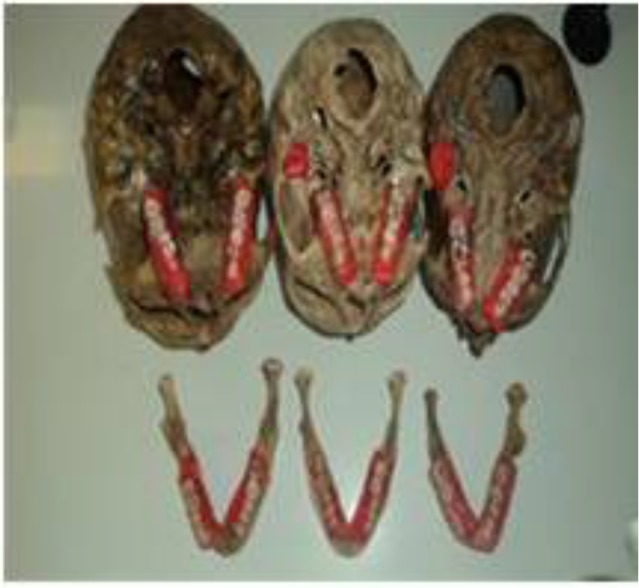
Materials and methods: In this in-vitro study, 100 teeth were placed in the dental sockets of a dry skull, and were fixed in normal proximal contacts. CBCT images were taken and were evaluated by two observers on the panoramic view at 1-, 3- and 5-mm-thick cross-sections, with the use of filtrations 0, 1 and 2. Afterwards, the samples were sectioned and underwent a histological evaluation. McNemar's test was used to compare the findings on CBCT images and histological evaluation. Receiver operating characteristic (ROC) curves and logistic regression were used to evaluate the diagnostic accuracy of different cross-sections.
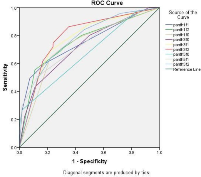
Results: The maximum AZ-value was achieved at 3-mm thickness/filtration 2. However, the differences between 1-mm thickness/filtration 2 and 1-mm thickness/filtration 1 were not significant (P=0.728 and 0.868, respectively). The minimum AZ-value was achieved at 5-mm thickness/filtration 0.
Conclusions: Although CBCT is not sufficiently effective in detecting caries, the best cross-sections for detection of proximal caries were achieved at 3-mm thickness/filtration 2, 1-mm thickness/filtration 2 and 1-mm thickness/filtration 1.

 求助内容:
求助内容: 应助结果提醒方式:


