{"title":"c-kit Positive Cardiac Outgrowth Cells Demonstrate Better Ability for Cardiac Recovery Against Ischemic Myopathy.","authors":"Chuan Li, Satoshi Matsushita, Zhengqing Li, Jianjun Guan, Atsushi Amano","doi":"10.4172/2157-7633.1000402","DOIUrl":null,"url":null,"abstract":"<p><strong>Objective: </strong>Resident cardiac stem cells are expected to be a therapeutic option for patients who suffer from severe heart failure. However, uncertainty remains over whether sorting cells for c-kit, a stem cell marker, improves therapeutic outcomes.</p><p><strong>Materials and methods: </strong>Cardiac outgrowth cells cultured from explants of rat heart atrium were sorted according to their positivity (+) or negativity (-) for c-kit. These cells were exposed to hypoxia for 3 d, and subsequently harvested for mRNA expression measurement. The cell medium was also collected to assess cytokine secretion. To test for a functional benefit in animals, myocardial infarction (MI) was induced in rats, and c-kit+ or c-kit- cells were injected. The left ventricular ejection fraction (LVEF) was measured for up to 4 weeks, after which the heart was harvested for biological and histological analyses.</p><p><strong>Results and conclusion: </strong>Expression of the angiogenesis-related genes, VEGF and ANGPTL2, was significantly higher in c-kit+ cells after 3 d of hypoxic culture, although we found no such difference prior to hypoxia. Secretion of VEGF and ANGPTL2 was greater in the c-kit+ group than in the c-kit- group, while hypoxia tended to increase cytokine expression in both groups. In addition, IGF-1 was significantly increased in the c-kit+ group, consistent with the relatively low expression of cleaved-caspase 3 revealed by western blot assay, and the relatively low count of apoptotic cells revealed by histochemical analysis. Administration of c-kit+cells into the MI heart improved the LVEF and increased neovascularization. These results indicate that c-kit+cells may be useful in cardiac stem cell therapy.</p>","PeriodicalId":89694,"journal":{"name":"Journal of stem cell research & therapy","volume":"7 10","pages":""},"PeriodicalIF":0.0000,"publicationDate":"2017-10-01","publicationTypes":"Journal Article","fieldsOfStudy":null,"isOpenAccess":false,"openAccessPdf":"https://sci-hub-pdf.com/10.4172/2157-7633.1000402","citationCount":"9","resultStr":null,"platform":"Semanticscholar","paperid":null,"PeriodicalName":"Journal of stem cell research & therapy","FirstCategoryId":"1085","ListUrlMain":"https://doi.org/10.4172/2157-7633.1000402","RegionNum":0,"RegionCategory":null,"ArticlePicture":[],"TitleCN":null,"AbstractTextCN":null,"PMCID":null,"EPubDate":"2017/10/13 0:00:00","PubModel":"Epub","JCR":"","JCRName":"","Score":null,"Total":0}
引用次数: 9
Abstract
Objective: Resident cardiac stem cells are expected to be a therapeutic option for patients who suffer from severe heart failure. However, uncertainty remains over whether sorting cells for c-kit, a stem cell marker, improves therapeutic outcomes.
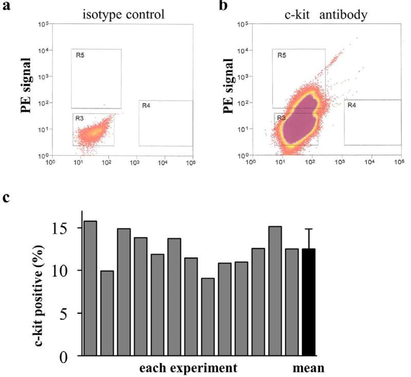
Materials and methods: Cardiac outgrowth cells cultured from explants of rat heart atrium were sorted according to their positivity (+) or negativity (-) for c-kit. These cells were exposed to hypoxia for 3 d, and subsequently harvested for mRNA expression measurement. The cell medium was also collected to assess cytokine secretion. To test for a functional benefit in animals, myocardial infarction (MI) was induced in rats, and c-kit+ or c-kit- cells were injected. The left ventricular ejection fraction (LVEF) was measured for up to 4 weeks, after which the heart was harvested for biological and histological analyses.
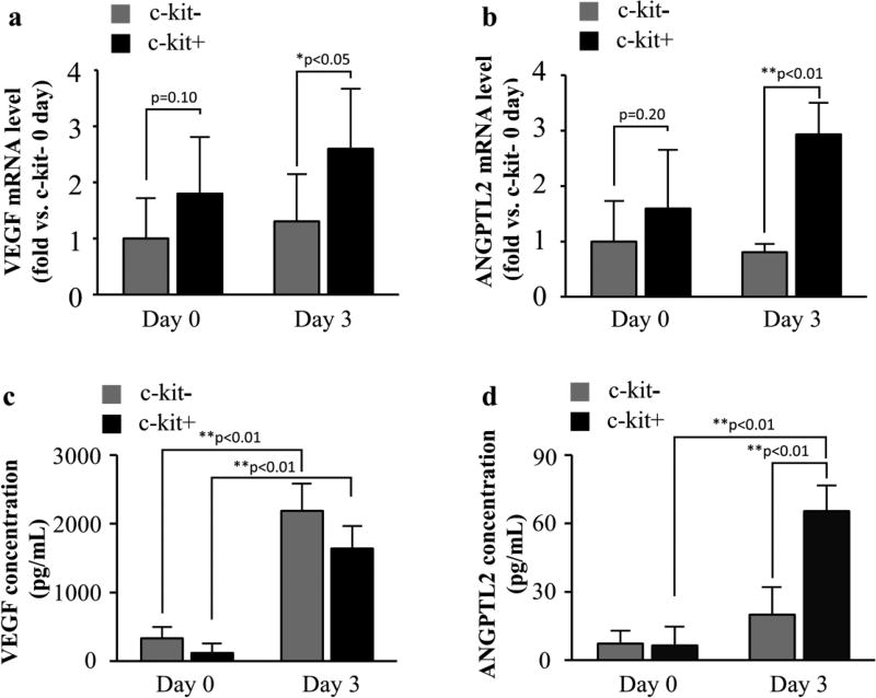
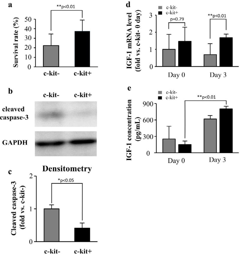
Results and conclusion: Expression of the angiogenesis-related genes, VEGF and ANGPTL2, was significantly higher in c-kit+ cells after 3 d of hypoxic culture, although we found no such difference prior to hypoxia. Secretion of VEGF and ANGPTL2 was greater in the c-kit+ group than in the c-kit- group, while hypoxia tended to increase cytokine expression in both groups. In addition, IGF-1 was significantly increased in the c-kit+ group, consistent with the relatively low expression of cleaved-caspase 3 revealed by western blot assay, and the relatively low count of apoptotic cells revealed by histochemical analysis. Administration of c-kit+cells into the MI heart improved the LVEF and increased neovascularization. These results indicate that c-kit+cells may be useful in cardiac stem cell therapy.

 求助内容:
求助内容: 应助结果提醒方式:


