{"title":"Posterior Epidural Herniation of a Lumbar Disk Fragment at L2-3 That Mimicked an Epidural Hematoma.","authors":"Jin-Sang Kil, Jong-Tae Park","doi":"10.14245/kjs.2017.14.3.115","DOIUrl":null,"url":null,"abstract":"<p><p>Lumbar disk herniation is common. Because of the posterior longitudinal ligament, migration usually occurs into the ventral epidural space. Rarely, fragments migrate into the dorsal epidural space. A 57-year-old man presented with lower back pain and weakness on right hip flexion and right knee flexion. He had lower back pain 1 day previously and received a transforaminal epidural block at a local hospital. The next day, he reported weakness of the right lower extremity. Lumbar spine magnetic resonance imaging revealed a dorsal epidural lesion with compression of the thecal sac at L2-3. Initial differential diagnoses included epidural hematoma after the block, neoplasm, and a sequestrated disk. Posterior lumbar decompression was performed. The lesion was identified intraoperatively as a large herniated disk fragment. Posterior epidural herniation of a lumbar disk fragment is rare and may be difficult to diagnose preoperatively. It may present as a variety of clinical scenarios and, as in this case, may mimic epidural hematoma.</p>","PeriodicalId":17867,"journal":{"name":"Korean Journal of Spine","volume":"14 3","pages":"115-117"},"PeriodicalIF":0.0000,"publicationDate":"2017-09-01","publicationTypes":"Journal Article","fieldsOfStudy":null,"isOpenAccess":false,"openAccessPdf":"https://ftp.ncbi.nlm.nih.gov/pub/pmc/oa_pdf/2b/9a/kjs-14-3-115.PMC5642095.pdf","citationCount":"11","resultStr":null,"platform":"Semanticscholar","paperid":null,"PeriodicalName":"Korean Journal of Spine","FirstCategoryId":"1085","ListUrlMain":"https://doi.org/10.14245/kjs.2017.14.3.115","RegionNum":0,"RegionCategory":null,"ArticlePicture":[],"TitleCN":null,"AbstractTextCN":null,"PMCID":null,"EPubDate":"2017/9/30 0:00:00","PubModel":"Epub","JCR":"","JCRName":"","Score":null,"Total":0}
引用次数: 11
Abstract
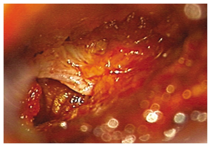
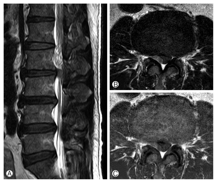
Lumbar disk herniation is common. Because of the posterior longitudinal ligament, migration usually occurs into the ventral epidural space. Rarely, fragments migrate into the dorsal epidural space. A 57-year-old man presented with lower back pain and weakness on right hip flexion and right knee flexion. He had lower back pain 1 day previously and received a transforaminal epidural block at a local hospital. The next day, he reported weakness of the right lower extremity. Lumbar spine magnetic resonance imaging revealed a dorsal epidural lesion with compression of the thecal sac at L2-3. Initial differential diagnoses included epidural hematoma after the block, neoplasm, and a sequestrated disk. Posterior lumbar decompression was performed. The lesion was identified intraoperatively as a large herniated disk fragment. Posterior epidural herniation of a lumbar disk fragment is rare and may be difficult to diagnose preoperatively. It may present as a variety of clinical scenarios and, as in this case, may mimic epidural hematoma.

 求助内容:
求助内容: 应助结果提醒方式:


