Ju Hyung Lee, Sung Han Oh, Pyung Goo Cho, Eun Mi Han, Je Beom Hong
{"title":"Solitary Osteochondroma Presenting as a Dumbbell Tumor Compressing the Cervical Spinal Cord.","authors":"Ju Hyung Lee, Sung Han Oh, Pyung Goo Cho, Eun Mi Han, Je Beom Hong","doi":"10.14245/kjs.2017.14.3.99","DOIUrl":null,"url":null,"abstract":"<p><p>We report a case of a solitary osteochondroma as a dumbbell tumor compressing the spinal cord and its surgical strategy. The patient is a 16-year-old female with longstanding posterior neck pain and left arm abduction weakness. She was examined by plain X-ray, three-dimensional-computed tomography, magnetic resonance imaging, and vertebral angiography. The analyses indicated a calcified extradural mass compressing the cord in the C3-4 portion extending into the neural and vertebral foramen with eroded vertebral body. The tumor was successfully excised using a modified combined anterior and posterior approach. Histopathologic study of the resected material confirmed the diagnosis. The postoperative assessment was followed by clinical and radiologically therapy for 5 years after surgery. Osteochondroma arises from enchondral bone but it rarely involves the spine, especially not as s dumbbell type. In this patient, the tumor may have arisen from the neural arch and extended into the extradural and extraforaminal space over a long period. We successfully removed the dumbbell tumor with a combined anterior oblique and posterior approach. However, further observation is essential because of the possibility of recurrence and sarcomatous change.</p>","PeriodicalId":17867,"journal":{"name":"Korean Journal of Spine","volume":"14 3","pages":"99-102"},"PeriodicalIF":0.0000,"publicationDate":"2017-09-01","publicationTypes":"Journal Article","fieldsOfStudy":null,"isOpenAccess":false,"openAccessPdf":"https://ftp.ncbi.nlm.nih.gov/pub/pmc/oa_pdf/73/88/kjs-14-3-99.PMC5642089.pdf","citationCount":"4","resultStr":null,"platform":"Semanticscholar","paperid":null,"PeriodicalName":"Korean Journal of Spine","FirstCategoryId":"1085","ListUrlMain":"https://doi.org/10.14245/kjs.2017.14.3.99","RegionNum":0,"RegionCategory":null,"ArticlePicture":[],"TitleCN":null,"AbstractTextCN":null,"PMCID":null,"EPubDate":"2017/9/30 0:00:00","PubModel":"Epub","JCR":"","JCRName":"","Score":null,"Total":0}
引用次数: 4
Abstract
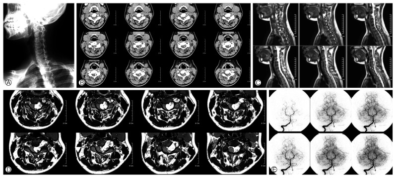
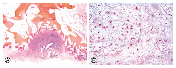
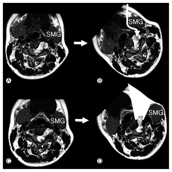
We report a case of a solitary osteochondroma as a dumbbell tumor compressing the spinal cord and its surgical strategy. The patient is a 16-year-old female with longstanding posterior neck pain and left arm abduction weakness. She was examined by plain X-ray, three-dimensional-computed tomography, magnetic resonance imaging, and vertebral angiography. The analyses indicated a calcified extradural mass compressing the cord in the C3-4 portion extending into the neural and vertebral foramen with eroded vertebral body. The tumor was successfully excised using a modified combined anterior and posterior approach. Histopathologic study of the resected material confirmed the diagnosis. The postoperative assessment was followed by clinical and radiologically therapy for 5 years after surgery. Osteochondroma arises from enchondral bone but it rarely involves the spine, especially not as s dumbbell type. In this patient, the tumor may have arisen from the neural arch and extended into the extradural and extraforaminal space over a long period. We successfully removed the dumbbell tumor with a combined anterior oblique and posterior approach. However, further observation is essential because of the possibility of recurrence and sarcomatous change.

 求助内容:
求助内容: 应助结果提醒方式:


