Treatment of a Four-Rooted Maxillary Second Molar Detected with Cone-Beam Computed Tomography.
Journal of Dentistry of Tehran University of Medical Sciences
Pub Date : 2017-03-01
引用次数: 0
Abstract
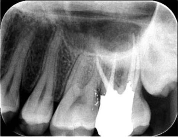
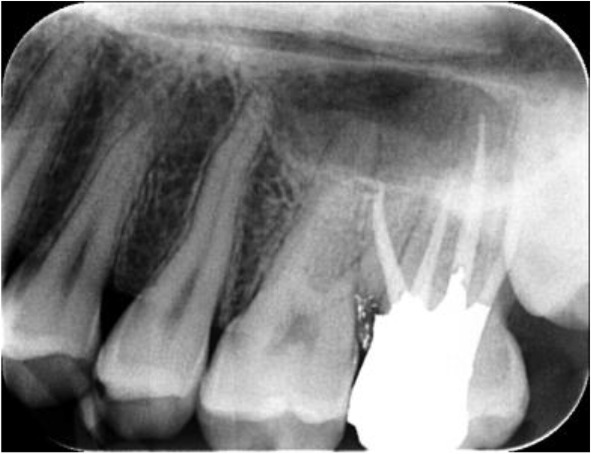
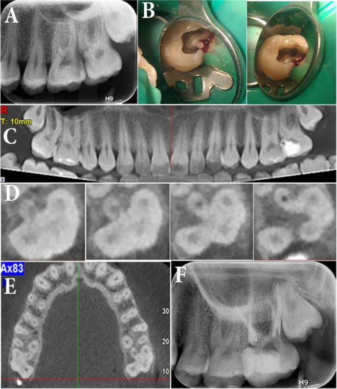
The significance of clinician's knowledge about root canal anatomy and its possible variations cannot be overlooked. In some cases, taking advantage of complementary imaging techniques can help achieve a perfect flawless endodontic treatment. This article reports endodontic management of a second maxillary molar that had an uncommon anatomy of the chamber floor. After obtaining a cone-beam computed tomography (CBCT) image, the presence of a second palatal root was confirmed. All four roots were treated and patient's symptoms were resolved.
锥束计算机断层扫描检测上颌四根第二磨牙的治疗。
临床医生对根管解剖及其可能变化的认识的重要性不容忽视。在某些情况下,利用互补成像技术可以帮助实现完美无瑕的牙髓治疗。这篇文章报告根管管理的第二上颌磨牙,有一个不常见的解剖室底。在获得锥形束计算机断层扫描(CBCT)图像后,证实了第二腭根的存在。所有四根均得到治疗,患者症状得到缓解。
本文章由计算机程序翻译,如有差异,请以英文原文为准。
求助全文
约1分钟内获得全文
求助全文

 求助内容:
求助内容: 应助结果提醒方式:


