{"title":"Investigation of impacted supernumerary teeth: a cone beam computed tomograph (cbct) study.","authors":"Gokhan Gurler, Cagri Delilbasi, Evren Delilbasi","doi":"10.17096/jiufd.20098","DOIUrl":null,"url":null,"abstract":"<p><strong>Purpose: </strong>The purpose of this study was to investigate the impacted supernumerary teeth which were initially detected on panoramic radiographs by using cone beam computed tomography (CBCT).</p><p><strong>Materials and methods: </strong>In this retrospective study, supernumerary teeth diagnosed on panoramic radiographs taken from patients who had admitted for routine dental treatment were evaluated using CBCT. Patients' age, gender, systemic conditions as well as number of supernumerary teeth, unilateral-bilateral presence, anatomical localization (maxilla, mandible, anterior-premolar-molar, mesiodens-lateral-canine, parapremolar-paramolar-distomolar) shape (rudimentary, supplemental, tuberculate, odontoma), position (palatal-lingual-buccal-labial-central), shortest distance between the tooth and adjacent cortical plate, complications and treatment were assessed.</p><p><strong>Results: </strong>A total of 47 impacted supernumerary teeth in 34 patients were investigated in this study. Of these, 33 (70.2%) were unilateral and 14 (29.8%) were bilateral. Only 1 supernumerary tooth was found in 27 patients (79.4%) whereas 7 patients (20.6%) had 2 or more supernumerary teeth. Most of the teeth located in the anterior region (74.4%) of the jaws and maxilla (74.4%). Twenty teeth (42.5%) were mesiodens, 11 (23.4%) were lateral or canine, 14 (29.7%) were parapremolar and 2(4.4%) were distomolar. Twenty-seven teeth (57.4%) were rudimentary, 15 (31.9%) were supplemental and 5 (10.7%) were odontoma in shape. The shortest distance between the supernumerary tooth and adjacent cortical plate varied between 0 to 2.5 mm with a mean of 0.66 mm. The most common clinical complaint was the non-eruption of permanent teeth (42.5%). All supernumerary teeth were removed under local anesthesia. Orthodontic traction was performed for those impacted permanent teeth if necessary.</p><p><strong>Conclusion: </strong>Impacted supernumerary teeth are usually in close proximity to cortical bone. Although this may facilitate surgical access, there is a risk of damaging surrounding anatomical structures. Therefore, CBCT evaluation of impacted supernumerary teeth for accurate planning is recommended.</p>","PeriodicalId":30947,"journal":{"name":"Journal of Istanbul University Faculty of Dentistry","volume":"51 3","pages":"18-24"},"PeriodicalIF":0.0000,"publicationDate":"2017-10-02","publicationTypes":"Journal Article","fieldsOfStudy":null,"isOpenAccess":false,"openAccessPdf":"https://sci-hub-pdf.com/10.17096/jiufd.20098","citationCount":"26","resultStr":null,"platform":"Semanticscholar","paperid":null,"PeriodicalName":"Journal of Istanbul University Faculty of Dentistry","FirstCategoryId":"1085","ListUrlMain":"https://doi.org/10.17096/jiufd.20098","RegionNum":0,"RegionCategory":null,"ArticlePicture":[],"TitleCN":null,"AbstractTextCN":null,"PMCID":null,"EPubDate":"2017/1/1 0:00:00","PubModel":"eCollection","JCR":"","JCRName":"","Score":null,"Total":0}
引用次数: 26
Abstract
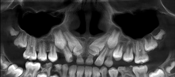
Purpose: The purpose of this study was to investigate the impacted supernumerary teeth which were initially detected on panoramic radiographs by using cone beam computed tomography (CBCT).
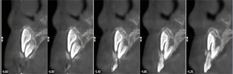
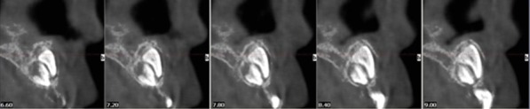
Materials and methods: In this retrospective study, supernumerary teeth diagnosed on panoramic radiographs taken from patients who had admitted for routine dental treatment were evaluated using CBCT. Patients' age, gender, systemic conditions as well as number of supernumerary teeth, unilateral-bilateral presence, anatomical localization (maxilla, mandible, anterior-premolar-molar, mesiodens-lateral-canine, parapremolar-paramolar-distomolar) shape (rudimentary, supplemental, tuberculate, odontoma), position (palatal-lingual-buccal-labial-central), shortest distance between the tooth and adjacent cortical plate, complications and treatment were assessed.
Results: A total of 47 impacted supernumerary teeth in 34 patients were investigated in this study. Of these, 33 (70.2%) were unilateral and 14 (29.8%) were bilateral. Only 1 supernumerary tooth was found in 27 patients (79.4%) whereas 7 patients (20.6%) had 2 or more supernumerary teeth. Most of the teeth located in the anterior region (74.4%) of the jaws and maxilla (74.4%). Twenty teeth (42.5%) were mesiodens, 11 (23.4%) were lateral or canine, 14 (29.7%) were parapremolar and 2(4.4%) were distomolar. Twenty-seven teeth (57.4%) were rudimentary, 15 (31.9%) were supplemental and 5 (10.7%) were odontoma in shape. The shortest distance between the supernumerary tooth and adjacent cortical plate varied between 0 to 2.5 mm with a mean of 0.66 mm. The most common clinical complaint was the non-eruption of permanent teeth (42.5%). All supernumerary teeth were removed under local anesthesia. Orthodontic traction was performed for those impacted permanent teeth if necessary.
Conclusion: Impacted supernumerary teeth are usually in close proximity to cortical bone. Although this may facilitate surgical access, there is a risk of damaging surrounding anatomical structures. Therefore, CBCT evaluation of impacted supernumerary teeth for accurate planning is recommended.

 求助内容:
求助内容: 应助结果提醒方式:


