{"title":"Diagnostic accuracy of cone-beam computed tomography in detecting secondary caries under composite fillings: an in vitro study.","authors":"Elif Yildizer Keris, Oguzhan Demirel, Melih Ozdede, Bulent Altunkaynak, Ilkay Peker","doi":"10.17096/jiufd.62563","DOIUrl":null,"url":null,"abstract":"<p><strong>Purpose: </strong>The aim of this in vitro study was to assess the diagnostic performance of cone-beam computed tomography (CBCT) in the detection of secondary carious lesions under composite resin fillings applied to different types of cavities.</p><p><strong>Materials and methods: </strong>Occlusal cavities (O) (n=18), occlusal cavities with mesial or distal component (MO/DO) (n=30), and mesial-occlusal-distal cavities (MOD) (n=30) were prepared in seventy eight extracted human posterior teeth. In half of the cavities in each group, artificial secondary caries lesions were simulated. All cavities were restored by using composite resin. All specimens were embedded in silicone and they were positioned to have approximal contacts. CBCT imaging was done and data were evaluated two times with two week interval by two observers, using a five-point confidence scale. Intra- and inter-observer agreements were calculated with Kappa statistics (κ). The area under (Az) the receiver operating characteristic (ROC) curve was used to evaluate the diagnostic accuracy.</p><p><strong>Results: </strong>Intra- (κ =0.89) and inter-observer (κ = 0.79) agreements were found to be excellent. Az values were highest for the O restorations which is followed by the MOD and DO/MO restorations. Az values for MOD and DO/MO restorations were very low and no statistically significant difference was found. Sensitivity for DO/MO restorations and specificity for MOD restorations were found to be the lowest values.</p><p><strong>Conclusion: </strong>Diagnostic performance of CBCT was higher in O composite restorations than MOD and DO/MO restorations for secondary caries detection. The use of alternative imaging methods rather than CBCT may be useful for evaluating secondary caries under composite MOD and DO/MO restorations.</p>","PeriodicalId":30947,"journal":{"name":"Journal of Istanbul University Faculty of Dentistry","volume":"51 1","pages":"22-27"},"PeriodicalIF":0.0000,"publicationDate":"2017-01-02","publicationTypes":"Journal Article","fieldsOfStudy":null,"isOpenAccess":false,"openAccessPdf":"https://sci-hub-pdf.com/10.17096/jiufd.62563","citationCount":"4","resultStr":null,"platform":"Semanticscholar","paperid":null,"PeriodicalName":"Journal of Istanbul University Faculty of Dentistry","FirstCategoryId":"1085","ListUrlMain":"https://doi.org/10.17096/jiufd.62563","RegionNum":0,"RegionCategory":null,"ArticlePicture":[],"TitleCN":null,"AbstractTextCN":null,"PMCID":null,"EPubDate":"2017/1/1 0:00:00","PubModel":"eCollection","JCR":"","JCRName":"","Score":null,"Total":0}
引用次数: 4
Abstract
Purpose: The aim of this in vitro study was to assess the diagnostic performance of cone-beam computed tomography (CBCT) in the detection of secondary carious lesions under composite resin fillings applied to different types of cavities.
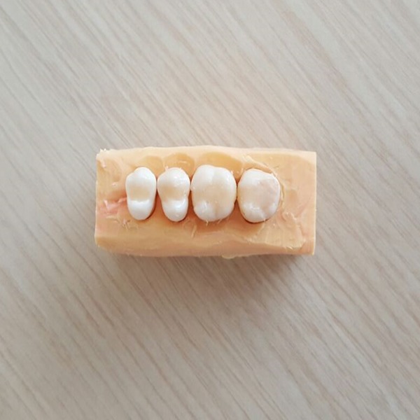
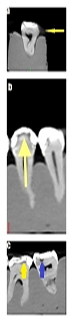
Materials and methods: Occlusal cavities (O) (n=18), occlusal cavities with mesial or distal component (MO/DO) (n=30), and mesial-occlusal-distal cavities (MOD) (n=30) were prepared in seventy eight extracted human posterior teeth. In half of the cavities in each group, artificial secondary caries lesions were simulated. All cavities were restored by using composite resin. All specimens were embedded in silicone and they were positioned to have approximal contacts. CBCT imaging was done and data were evaluated two times with two week interval by two observers, using a five-point confidence scale. Intra- and inter-observer agreements were calculated with Kappa statistics (κ). The area under (Az) the receiver operating characteristic (ROC) curve was used to evaluate the diagnostic accuracy.
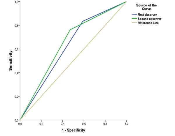
Results: Intra- (κ =0.89) and inter-observer (κ = 0.79) agreements were found to be excellent. Az values were highest for the O restorations which is followed by the MOD and DO/MO restorations. Az values for MOD and DO/MO restorations were very low and no statistically significant difference was found. Sensitivity for DO/MO restorations and specificity for MOD restorations were found to be the lowest values.
Conclusion: Diagnostic performance of CBCT was higher in O composite restorations than MOD and DO/MO restorations for secondary caries detection. The use of alternative imaging methods rather than CBCT may be useful for evaluating secondary caries under composite MOD and DO/MO restorations.

 求助内容:
求助内容: 应助结果提醒方式:


