{"title":"Magnetic Resonance-guided High Intensity Focused Ultrasound in the presence of biopsy markers.","authors":"Charles Mougenot, Chrit Moonen","doi":"10.1186/s40349-017-0103-1","DOIUrl":null,"url":null,"abstract":"<p><strong>Background: </strong>Magnetic Resonance guided High Intensity Focused ultrasound (MR-HIFU) offers precise non-invasive thermotherapy for clinical applications such as the treatment of breast lesions. However, patients with a biopsy marker are usually not eligible for MR-HIFU treatment. This study investigates the interaction of some MR-compatible markers with MR-HIFU thermotherapy.</p><p><strong>Methods: </strong>The MR-HIFU compatibility of 14 markers (6 Gold Anchor and 4 Visicoil markers in gold, 1 Visicoil marker in brass, 3 BiomarC markers in carbon coated) were tested using the Sonalleve breast MR-HIFU platform at 1.5 T. The impact of these markers was assessed by counting the number of voxels with low signal intensity on MR thermal maps and by comparing temperature increases induced by the HIFU beam.</p><p><strong>Results: </strong>Most markers were visible on thermal maps with an apparent size 4.2 ± 3.1 and 2 ± 1.8 times larger than their respective actual width and length. The volume of masked voxels was for most of the markers much larger than the actual volume of the marker (up to a factor 65.1). However, it represents only a small fraction of the 12 mm diameter targeted region (up to 8.8 voxels which represents 19% of this targeted region). Some differences in the maximal temperature increase were observed especially for BiomarC 1 × 3 and BiomarC 2 × 4 markers enhancing the heating. These differences were less pronounced at the edge of the targeted region.</p><p><strong>Conclusion: </strong>All markers had a minimal impact on the volume above the thermal dose threshold of 240 EM since the differences measured were smaller than the in-plane image resolution of 1.56 mm.</p>","PeriodicalId":90245,"journal":{"name":"Journal of therapeutic ultrasound","volume":"5 ","pages":"25"},"PeriodicalIF":0.0000,"publicationDate":"2017-09-20","publicationTypes":"Journal Article","fieldsOfStudy":null,"isOpenAccess":false,"openAccessPdf":"https://sci-hub-pdf.com/10.1186/s40349-017-0103-1","citationCount":"5","resultStr":null,"platform":"Semanticscholar","paperid":null,"PeriodicalName":"Journal of therapeutic ultrasound","FirstCategoryId":"1085","ListUrlMain":"https://doi.org/10.1186/s40349-017-0103-1","RegionNum":0,"RegionCategory":null,"ArticlePicture":[],"TitleCN":null,"AbstractTextCN":null,"PMCID":null,"EPubDate":"2017/1/1 0:00:00","PubModel":"eCollection","JCR":"","JCRName":"","Score":null,"Total":0}
引用次数: 5
Abstract
Background: Magnetic Resonance guided High Intensity Focused ultrasound (MR-HIFU) offers precise non-invasive thermotherapy for clinical applications such as the treatment of breast lesions. However, patients with a biopsy marker are usually not eligible for MR-HIFU treatment. This study investigates the interaction of some MR-compatible markers with MR-HIFU thermotherapy.
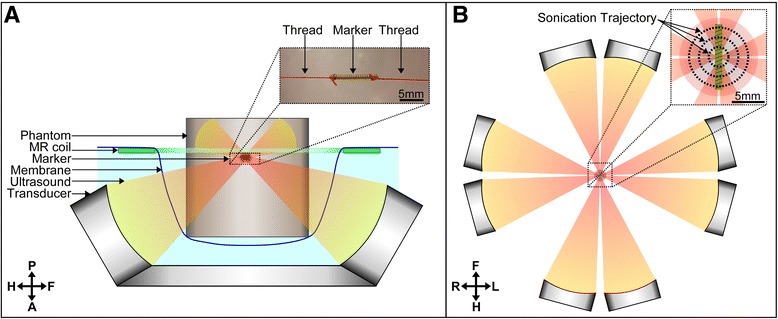
Methods: The MR-HIFU compatibility of 14 markers (6 Gold Anchor and 4 Visicoil markers in gold, 1 Visicoil marker in brass, 3 BiomarC markers in carbon coated) were tested using the Sonalleve breast MR-HIFU platform at 1.5 T. The impact of these markers was assessed by counting the number of voxels with low signal intensity on MR thermal maps and by comparing temperature increases induced by the HIFU beam.
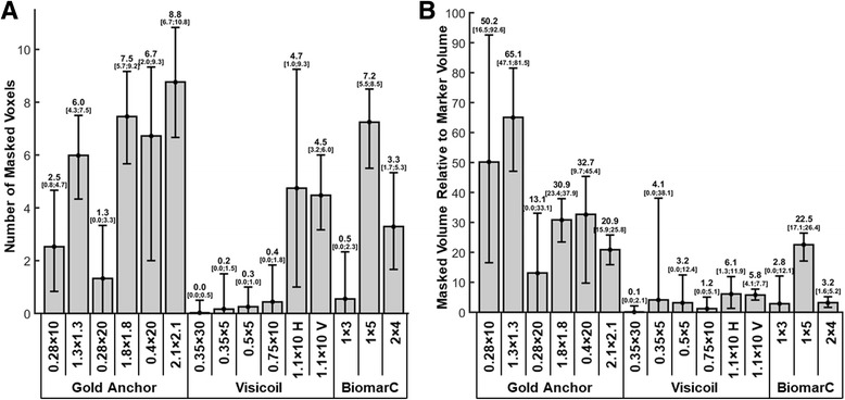
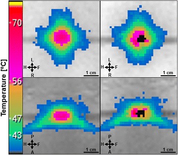
Results: Most markers were visible on thermal maps with an apparent size 4.2 ± 3.1 and 2 ± 1.8 times larger than their respective actual width and length. The volume of masked voxels was for most of the markers much larger than the actual volume of the marker (up to a factor 65.1). However, it represents only a small fraction of the 12 mm diameter targeted region (up to 8.8 voxels which represents 19% of this targeted region). Some differences in the maximal temperature increase were observed especially for BiomarC 1 × 3 and BiomarC 2 × 4 markers enhancing the heating. These differences were less pronounced at the edge of the targeted region.
Conclusion: All markers had a minimal impact on the volume above the thermal dose threshold of 240 EM since the differences measured were smaller than the in-plane image resolution of 1.56 mm.

 求助内容:
求助内容: 应助结果提醒方式:


