Comparative Analysis of Lipid Extracts and Imaging Mass Spectrometry for Evaluating Cerebral White Matter Biochemical Pathology in an Experimental Second-Hand Cigarette Smoke Exposure Model.
Alexander Krotow, Emine B Yalcin, Jared Kay, Suzanne M de la Monte
{"title":"Comparative Analysis of Lipid Extracts and Imaging Mass Spectrometry for Evaluating Cerebral White Matter Biochemical Pathology in an Experimental Second-Hand Cigarette Smoke Exposure Model.","authors":"Alexander Krotow, Emine B Yalcin, Jared Kay, Suzanne M de la Monte","doi":"10.4172/2469-9861.1000113","DOIUrl":null,"url":null,"abstract":"<p><strong>Background: </strong>White matter injury and degeneration are common features of developmental and aging-associated diseases, yet their pathobiological bases are poorly understood. However, recent advances in Matrix-Assisted Laser Desorption Ionization (MALDI) instruments and chemistry have provided critical tools for myelin-lipid analytical research.</p><p><strong>Design: </strong>This study characterizes Cigarette Smoke (CS) exposure effects on frontal lobe lipid ion profiles in adult male A/J mice that had been exposed to air for 8 weeks (A8), CS for 4 (CS4) or 8 weeks (CS8), or CS8 followed by 2 weeks recovery (CS8+R). MALDI data acquired by analysis of lipid extracts plated onto a ground steel target (high through-put) were compared with Imaging Mass Spectrometry (IMS).</p><p><strong>Results: </strong>MALDI-time-of-flight (TOF) detected 120 lipid ions with m/z's of 600 to 1300 (phospholipids and sulfatides) in samples plated onto the steel target or analyzed by IMS, but just 25 ions (18%) were detected by both methods. IMS more effectively detected ions in the highest m/z range, whereas the extracts had abundant middle-range m/z ions. The experimental groups were better discriminated by PCA and R-generated heat map hierarchical clustering of IMS data than lipid extract data. On the other hand, both methods clearly delineated the CS4, CS8 and CS8+R experimental groups from control.</p><p><strong>Conclusions: </strong>MALDI analysis of brain lipid extracts plated onto a ground steel target for high through-put studies, or imaged directly in tissue can be used to assess biochemical pathology of white matter neurodegeneration and responses to treatment.</p>","PeriodicalId":92098,"journal":{"name":"Mass spectrometry & purification techniques","volume":"2 1","pages":""},"PeriodicalIF":0.0000,"publicationDate":"2016-01-01","publicationTypes":"Journal Article","fieldsOfStudy":null,"isOpenAccess":false,"openAccessPdf":"https://sci-hub-pdf.com/10.4172/2469-9861.1000113","citationCount":"2","resultStr":null,"platform":"Semanticscholar","paperid":null,"PeriodicalName":"Mass spectrometry & purification techniques","FirstCategoryId":"1085","ListUrlMain":"https://doi.org/10.4172/2469-9861.1000113","RegionNum":0,"RegionCategory":null,"ArticlePicture":[],"TitleCN":null,"AbstractTextCN":null,"PMCID":null,"EPubDate":"2016/4/20 0:00:00","PubModel":"Epub","JCR":"","JCRName":"","Score":null,"Total":0}
引用次数: 2
Abstract
Background: White matter injury and degeneration are common features of developmental and aging-associated diseases, yet their pathobiological bases are poorly understood. However, recent advances in Matrix-Assisted Laser Desorption Ionization (MALDI) instruments and chemistry have provided critical tools for myelin-lipid analytical research.
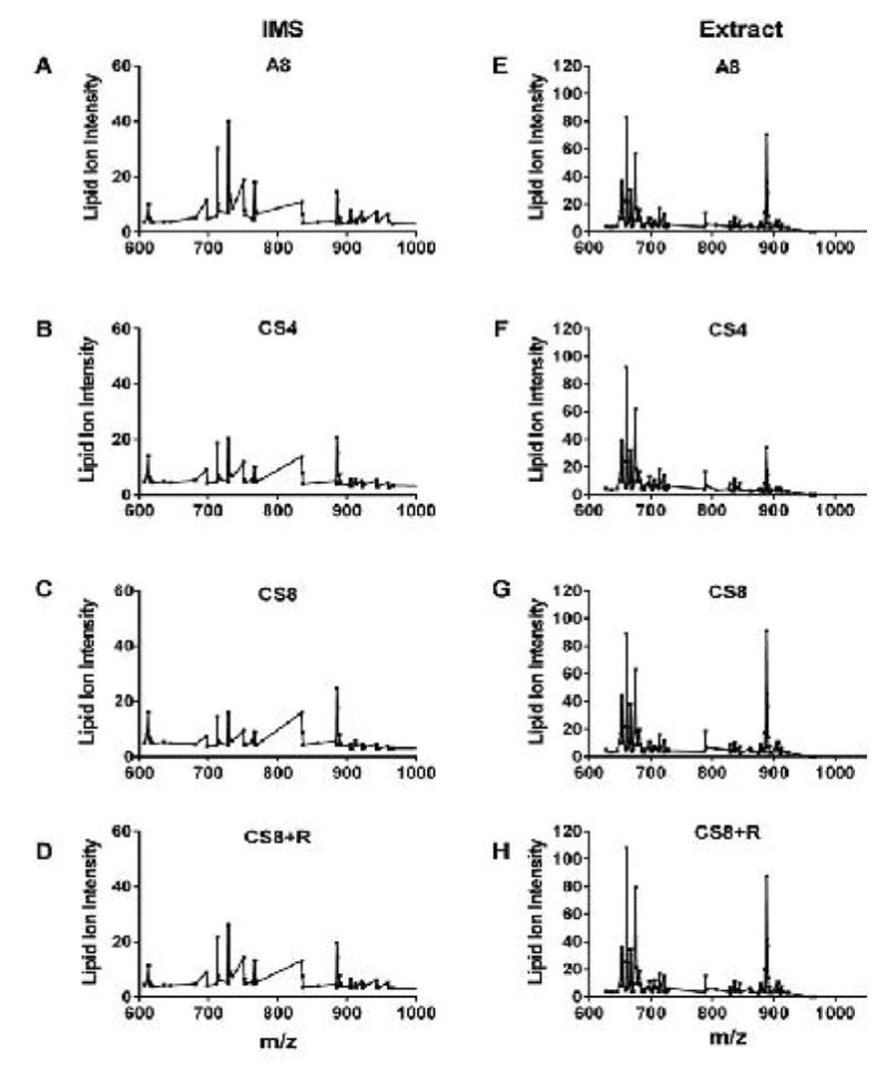
Design: This study characterizes Cigarette Smoke (CS) exposure effects on frontal lobe lipid ion profiles in adult male A/J mice that had been exposed to air for 8 weeks (A8), CS for 4 (CS4) or 8 weeks (CS8), or CS8 followed by 2 weeks recovery (CS8+R). MALDI data acquired by analysis of lipid extracts plated onto a ground steel target (high through-put) were compared with Imaging Mass Spectrometry (IMS).
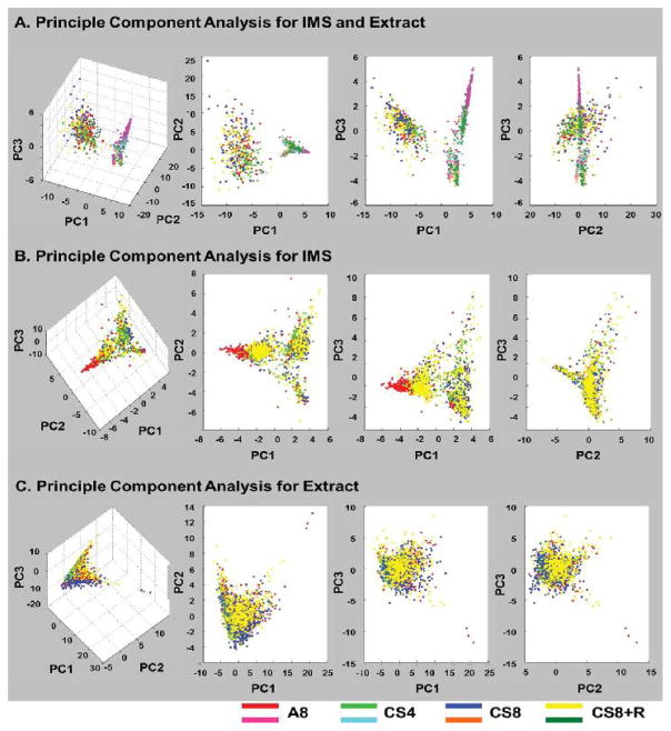
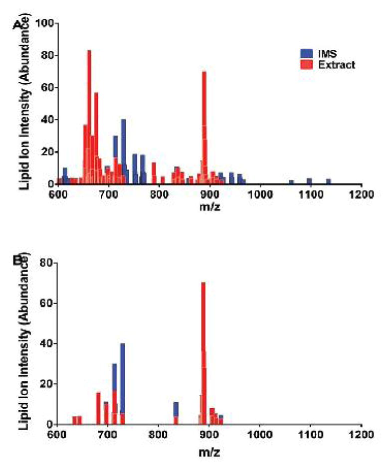
Results: MALDI-time-of-flight (TOF) detected 120 lipid ions with m/z's of 600 to 1300 (phospholipids and sulfatides) in samples plated onto the steel target or analyzed by IMS, but just 25 ions (18%) were detected by both methods. IMS more effectively detected ions in the highest m/z range, whereas the extracts had abundant middle-range m/z ions. The experimental groups were better discriminated by PCA and R-generated heat map hierarchical clustering of IMS data than lipid extract data. On the other hand, both methods clearly delineated the CS4, CS8 and CS8+R experimental groups from control.
Conclusions: MALDI analysis of brain lipid extracts plated onto a ground steel target for high through-put studies, or imaged directly in tissue can be used to assess biochemical pathology of white matter neurodegeneration and responses to treatment.

 求助内容:
求助内容: 应助结果提醒方式:


