Taeshin Kim, Bum-Joon Kim, Se-Hoon Kim, Seung-Hwan Lee
{"title":"Tophaceous Gout in the Lumbar Spinal Canal Mimicking Epidural Spinal Tumor.","authors":"Taeshin Kim, Bum-Joon Kim, Se-Hoon Kim, Seung-Hwan Lee","doi":"10.14245/kjs.2017.14.2.50","DOIUrl":null,"url":null,"abstract":"<p><p>Gout is an inflammatory arthritis characterized by deposition of monosodium urate crystals in joints. Though gout frequently involves the big toe or other extremities, it rarely occurs in the spinal canal. A 35-year-old man presented with left L5 radiculopathy. He had leg pain for 8 months and received several epidural steroid injections. Magnetic resonance imaging revealed a 1.7×1.1-cm ovoid contrast-enhancing mass, causing pressure erosion of the left L5 pedicle. Microscopic laminotomy was performed at the left L5 lamina. White chalky materials, identified at the left lateral recess of the spinal canal, were removed in a piecemeal manner. The histopathologic diagnosis was tophaceous gout. Although the patient's radiating pain did not resolve postoperatively, it was dramatically relieved with uric acid-lowering medications. If a mass effect is suspected, surgical removal of gouty tophi might aid in symptom release and definite diagnosis. Medical treatment after rheumatology consultation is crucial.</p>","PeriodicalId":17867,"journal":{"name":"Korean Journal of Spine","volume":"14 2","pages":"50-52"},"PeriodicalIF":0.0000,"publicationDate":"2017-06-01","publicationTypes":"Journal Article","fieldsOfStudy":null,"isOpenAccess":false,"openAccessPdf":"https://ftp.ncbi.nlm.nih.gov/pub/pmc/oa_pdf/f6/9c/kjs-14-2-50.PMC5518431.pdf","citationCount":"8","resultStr":null,"platform":"Semanticscholar","paperid":null,"PeriodicalName":"Korean Journal of Spine","FirstCategoryId":"1085","ListUrlMain":"https://doi.org/10.14245/kjs.2017.14.2.50","RegionNum":0,"RegionCategory":null,"ArticlePicture":[],"TitleCN":null,"AbstractTextCN":null,"PMCID":null,"EPubDate":"2017/6/30 0:00:00","PubModel":"Epub","JCR":"","JCRName":"","Score":null,"Total":0}
引用次数: 8
Abstract
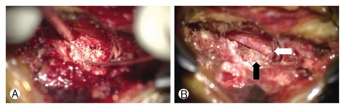
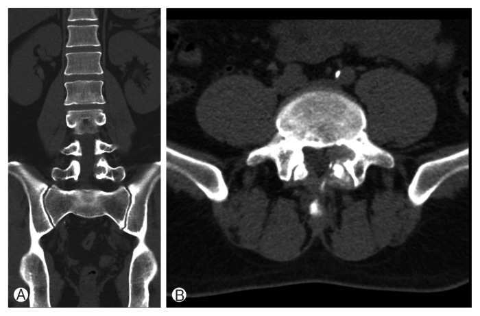
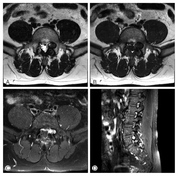
Gout is an inflammatory arthritis characterized by deposition of monosodium urate crystals in joints. Though gout frequently involves the big toe or other extremities, it rarely occurs in the spinal canal. A 35-year-old man presented with left L5 radiculopathy. He had leg pain for 8 months and received several epidural steroid injections. Magnetic resonance imaging revealed a 1.7×1.1-cm ovoid contrast-enhancing mass, causing pressure erosion of the left L5 pedicle. Microscopic laminotomy was performed at the left L5 lamina. White chalky materials, identified at the left lateral recess of the spinal canal, were removed in a piecemeal manner. The histopathologic diagnosis was tophaceous gout. Although the patient's radiating pain did not resolve postoperatively, it was dramatically relieved with uric acid-lowering medications. If a mass effect is suspected, surgical removal of gouty tophi might aid in symptom release and definite diagnosis. Medical treatment after rheumatology consultation is crucial.

 求助内容:
求助内容: 应助结果提醒方式:


