Anne K Maxwell, Henry P Barham, Anne E Getz, Todd T Kingdom, Vijay R Ramakrishnan
{"title":"Landmarks for rapid localization of the sphenopalatine foramen: A radiographic morphometric analysis.","authors":"Anne K Maxwell, Henry P Barham, Anne E Getz, Todd T Kingdom, Vijay R Ramakrishnan","doi":"10.2500/ar.2017.8.0196","DOIUrl":null,"url":null,"abstract":"<p><strong>Background: </strong>Transnasal endoscopic sphenopalatine artery ligation is becoming the procedure of choice for surgical management of intractable posterior epistaxis. Landmarks for localization of the sphenopalatine foramen can assist in rapid surgical exposure of the sphenopalatine artery.</p><p><strong>Objective: </strong>This study examined distances from easily identified endoscopic surgical landmarks to the sphenopalatine foramen.</p><p><strong>Methods: </strong>By using computed tomography of the sinus to study radiologic anatomy in 50 adults, distances were measured between five simple endoscopic landmarks and the sphenopalatine foramen. The two-tailed t-test was used for statistical analysis.</p><p><strong>Results: </strong>Right- and left-sided measurements were similar. The mean (standard deviation [SD]) anteroposterior distances to the sphenopalatine foramen were the following: from the maxillary line (36.7 ± 5.5 mm), anterior head of the middle turbinate (33.8 ± 6.7 mm), basal lamella (11.8 ± 1.9 mm), and choanal arch (-9.2 ± 1.4 mm). The mean (SD) distance in the vertical dimension from the nasal floor was 26.6 ± 2.6 mm. Female patients had statistically shorter distances to the sphenopalatine foramen from the maxillary line, anterior head of the middle turbinate, choanal arch, and nasal floor.</p><p><strong>Conclusion: </strong>Reliable endoscopic landmarks exist in relation to consistent anatomic structures and can be used to help quickly estimate the location of the sphenopalatine foramen at the onset of the procedure.</p>","PeriodicalId":45192,"journal":{"name":"Allergy & Rhinology","volume":"8 2","pages":"63-66"},"PeriodicalIF":2.3000,"publicationDate":"2017-06-01","publicationTypes":"Journal Article","fieldsOfStudy":null,"isOpenAccess":false,"openAccessPdf":"https://sci-hub-pdf.com/10.2500/ar.2017.8.0196","citationCount":"7","resultStr":null,"platform":"Semanticscholar","paperid":null,"PeriodicalName":"Allergy & Rhinology","FirstCategoryId":"1085","ListUrlMain":"https://doi.org/10.2500/ar.2017.8.0196","RegionNum":0,"RegionCategory":null,"ArticlePicture":[],"TitleCN":null,"AbstractTextCN":null,"PMCID":null,"EPubDate":"","PubModel":"","JCR":"Q1","JCRName":"OTORHINOLARYNGOLOGY","Score":null,"Total":0}
引用次数: 7
Abstract
Background: Transnasal endoscopic sphenopalatine artery ligation is becoming the procedure of choice for surgical management of intractable posterior epistaxis. Landmarks for localization of the sphenopalatine foramen can assist in rapid surgical exposure of the sphenopalatine artery.
Objective: This study examined distances from easily identified endoscopic surgical landmarks to the sphenopalatine foramen.
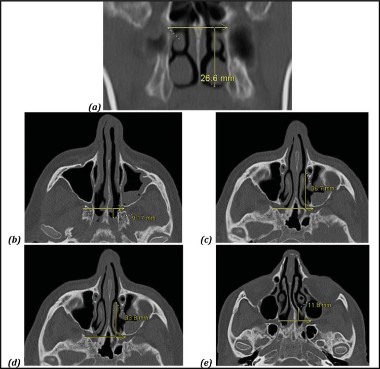
Methods: By using computed tomography of the sinus to study radiologic anatomy in 50 adults, distances were measured between five simple endoscopic landmarks and the sphenopalatine foramen. The two-tailed t-test was used for statistical analysis.
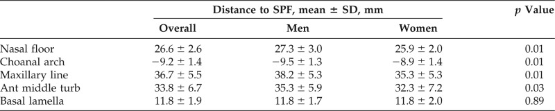
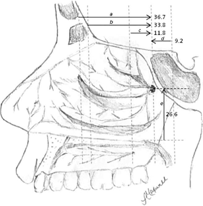
Results: Right- and left-sided measurements were similar. The mean (standard deviation [SD]) anteroposterior distances to the sphenopalatine foramen were the following: from the maxillary line (36.7 ± 5.5 mm), anterior head of the middle turbinate (33.8 ± 6.7 mm), basal lamella (11.8 ± 1.9 mm), and choanal arch (-9.2 ± 1.4 mm). The mean (SD) distance in the vertical dimension from the nasal floor was 26.6 ± 2.6 mm. Female patients had statistically shorter distances to the sphenopalatine foramen from the maxillary line, anterior head of the middle turbinate, choanal arch, and nasal floor.
Conclusion: Reliable endoscopic landmarks exist in relation to consistent anatomic structures and can be used to help quickly estimate the location of the sphenopalatine foramen at the onset of the procedure.

 求助内容:
求助内容: 应助结果提醒方式:


