Yu Meng, Changzhen Shi, Bo Hu, Jian Gong, Xing Zhong, Xueyin Lin, Xinju Zhang, Jun Liu, Cong Liu, Hao Xu
{"title":"External magnetic field promotes homing of magnetized stem cells following subcutaneous injection.","authors":"Yu Meng, Changzhen Shi, Bo Hu, Jian Gong, Xing Zhong, Xueyin Lin, Xinju Zhang, Jun Liu, Cong Liu, Hao Xu","doi":"10.1186/s12860-017-0140-1","DOIUrl":null,"url":null,"abstract":"<p><strong>Background: </strong>Mesenchymal stem cells (MSCs) are multipotent stromal cells that have the ability to self-renew and migrate to sites of pathology. In vivo tracking of MSCs provides insights into both, the underlying mechanisms of MSC transformation and their potential as gene delivery vehicles. The aim of our study was to assess the ability of superparamagnetic iron oxide nanoparticles (SPIONs)-labeled Wharton's Jelly of the human umbilical cord-derived MSCs (WJ-MSCs) to carry the green fluorescent protein (GFP) gene to cutaneous injury sites in a murine model.</p><p><strong>Methods: </strong>WJ-MSCs were isolated from a fresh umbilical cord and were genetically transformed to carry the GFP gene using lentiviral vectors with magnetically labeled SPIONs. The SPIONs/GFP-positive WJ-MSCs expressed multipotent cell markers and demonstrated the potential for osteogenic and adipogenic differentiation. Fifteen skin-injured mice were divided into three groups. Group I was treated with WJ-MSCs, group II with SPIONs/GFP-positive WJ-MSCs, and group III with SPIONs/GFP-positive WJ-MSCs exposed to an external magnetic field (EMF). Magnetic resonance imaging and optical molecular imaging were performed, and images were acquired 1, 2, and 7 days after cell injection.</p><p><strong>Results: </strong>The results showed that GFP could be intensively detected around the wound in vivo 24 h after the cells were injected. Furthermore, we observed an accumulation of WJ-MSCs at the wound site, and EMF exposure increased the speed of cell transport. In conclusion, our study demonstrated that SPIONs/GFP function as cellular probes for monitoring in vivo migration and homing of WJ-MSCs. Moreover, exposure to an EMF can increase the transportation efficiency of SPIONs-labeled WJ-MSCs in vivo.</p><p><strong>Conclusions: </strong>Our findings could lead to the development of a gene carrier system for the treatment of diseases.</p>","PeriodicalId":9051,"journal":{"name":"BMC Cell Biology","volume":"18 1","pages":"24"},"PeriodicalIF":0.0000,"publicationDate":"2017-05-26","publicationTypes":"Journal Article","fieldsOfStudy":null,"isOpenAccess":false,"openAccessPdf":"https://sci-hub-pdf.com/10.1186/s12860-017-0140-1","citationCount":"1","resultStr":null,"platform":"Semanticscholar","paperid":null,"PeriodicalName":"BMC Cell Biology","FirstCategoryId":"1085","ListUrlMain":"https://doi.org/10.1186/s12860-017-0140-1","RegionNum":0,"RegionCategory":null,"ArticlePicture":[],"TitleCN":null,"AbstractTextCN":null,"PMCID":null,"EPubDate":"","PubModel":"","JCR":"Q1","JCRName":"Biochemistry, Genetics and Molecular Biology","Score":null,"Total":0}
引用次数: 1
Abstract
Background: Mesenchymal stem cells (MSCs) are multipotent stromal cells that have the ability to self-renew and migrate to sites of pathology. In vivo tracking of MSCs provides insights into both, the underlying mechanisms of MSC transformation and their potential as gene delivery vehicles. The aim of our study was to assess the ability of superparamagnetic iron oxide nanoparticles (SPIONs)-labeled Wharton's Jelly of the human umbilical cord-derived MSCs (WJ-MSCs) to carry the green fluorescent protein (GFP) gene to cutaneous injury sites in a murine model.
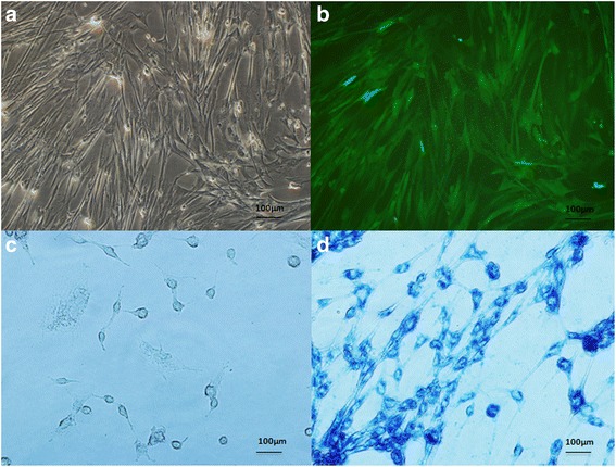
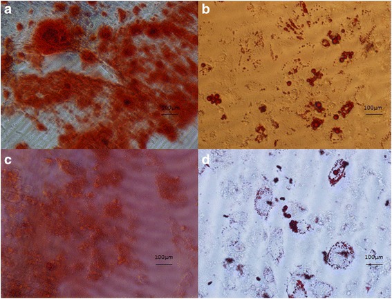
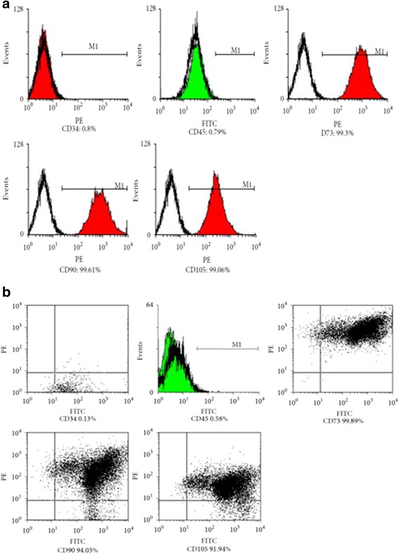
Methods: WJ-MSCs were isolated from a fresh umbilical cord and were genetically transformed to carry the GFP gene using lentiviral vectors with magnetically labeled SPIONs. The SPIONs/GFP-positive WJ-MSCs expressed multipotent cell markers and demonstrated the potential for osteogenic and adipogenic differentiation. Fifteen skin-injured mice were divided into three groups. Group I was treated with WJ-MSCs, group II with SPIONs/GFP-positive WJ-MSCs, and group III with SPIONs/GFP-positive WJ-MSCs exposed to an external magnetic field (EMF). Magnetic resonance imaging and optical molecular imaging were performed, and images were acquired 1, 2, and 7 days after cell injection.
Results: The results showed that GFP could be intensively detected around the wound in vivo 24 h after the cells were injected. Furthermore, we observed an accumulation of WJ-MSCs at the wound site, and EMF exposure increased the speed of cell transport. In conclusion, our study demonstrated that SPIONs/GFP function as cellular probes for monitoring in vivo migration and homing of WJ-MSCs. Moreover, exposure to an EMF can increase the transportation efficiency of SPIONs-labeled WJ-MSCs in vivo.
Conclusions: Our findings could lead to the development of a gene carrier system for the treatment of diseases.
期刊介绍:
BMC Molecular and Cell Biology, formerly known as BMC Cell Biology, is an open access journal that considers articles on all aspects of both eukaryotic and prokaryotic cell and molecular biology, including structural and functional cell biology, DNA and RNA in a cellular context and biochemistry, as well as research using both the experimental and theoretical aspects of physics to study biological processes and investigations into the structure of biological macromolecules.

 求助内容:
求助内容: 应助结果提醒方式:


