Correlation Between Ischemic Retinal Accidents and Radial Peripapillary Capillaries in the Optic Nerve Using Optical Coherence Tomographic Angiography: Observations in 6 Patients.

Ophthalmology and eye diseases Pub Date : 2017-04-17 eCollection Date: 2017-01-01 DOI:10.1177/1179172117702889
Marta Cerdà-Ibáñez, Antonio Duch-Samper, Rodrigo Clemente-Tomás, Raúl Torrecillas-Picazo, Noemí Ruiz Del Río, Laura Manfreda-Dominguez
{"title":"Correlation Between Ischemic Retinal Accidents and Radial Peripapillary Capillaries in the Optic Nerve Using Optical Coherence Tomographic Angiography: Observations in 6 Patients.","authors":"Marta Cerdà-Ibáñez, Antonio Duch-Samper, Rodrigo Clemente-Tomás, Raúl Torrecillas-Picazo, Noemí Ruiz Del Río, Laura Manfreda-Dominguez","doi":"10.1177/1179172117702889","DOIUrl":null,"url":null,"abstract":"Background: Perfusion of the optic nerve has been widely studied using fluorescein angiography (FAG), which is currently regarded as the criterion standard. However, FAG has adverse effects associated with intravenous contrast administration and is limited in its capacity to characterize and stratify the different vascular layers of the optic nerve and retina. The use of new imaging techniques, such as optical coherence tomographic angiography (Angio-OCT), is therefore important. Aim: A qualitative description is made of the vascular layers of the optic nerve and of how vascular events affect radial peripapillary capillaries (RPC). Two patients with central retinal artery occlusion (CRAO), 1 with arteritic anterior ischemic optic neuropathy (AAION), and 3 healthy subjects were studied. Results: The Angio-OCT imaging afforded better visualization of the depth of the RPC and rest of the vascular layers of the retina compared with FAG. Optic nerve surface perfusion was affected in AAION and proved normal in CRAO. Conclusions: Our results indicate that perfusion of the papilla and RPC mainly arises from the papillary plexus that depends on the posterior ciliary artery.","PeriodicalId":74362,"journal":{"name":"Ophthalmology and eye diseases","volume":" ","pages":"1179172117702889"},"PeriodicalIF":0.0000,"publicationDate":"2017-04-17","publicationTypes":"Journal Article","fieldsOfStudy":null,"isOpenAccess":false,"openAccessPdf":"https://sci-hub-pdf.com/10.1177/1179172117702889","citationCount":"9","resultStr":null,"platform":"Semanticscholar","paperid":null,"PeriodicalName":"Ophthalmology and eye diseases","FirstCategoryId":"1085","ListUrlMain":"https://doi.org/10.1177/1179172117702889","RegionNum":0,"RegionCategory":null,"ArticlePicture":[],"TitleCN":null,"AbstractTextCN":null,"PMCID":null,"EPubDate":"2017/1/1 0:00:00","PubModel":"eCollection","JCR":"","JCRName":"","Score":null,"Total":0}
引用次数: 9

Abstract

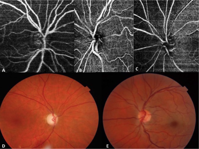
Background: Perfusion of the optic nerve has been widely studied using fluorescein angiography (FAG), which is currently regarded as the criterion standard. However, FAG has adverse effects associated with intravenous contrast administration and is limited in its capacity to characterize and stratify the different vascular layers of the optic nerve and retina. The use of new imaging techniques, such as optical coherence tomographic angiography (Angio-OCT), is therefore important. Aim: A qualitative description is made of the vascular layers of the optic nerve and of how vascular events affect radial peripapillary capillaries (RPC). Two patients with central retinal artery occlusion (CRAO), 1 with arteritic anterior ischemic optic neuropathy (AAION), and 3 healthy subjects were studied. Results: The Angio-OCT imaging afforded better visualization of the depth of the RPC and rest of the vascular layers of the retina compared with FAG. Optic nerve surface perfusion was affected in AAION and proved normal in CRAO. Conclusions: Our results indicate that perfusion of the papilla and RPC mainly arises from the papillary plexus that depends on the posterior ciliary artery.
Abstract Image

光学相干层析血管成像与视神经径向乳头周围毛细血管的相关性:6例观察。
背景:视神经灌注的荧光素血管造影(FAG)已被广泛研究,目前被视为标准。然而,FAG与静脉注射造影剂相关的不良反应,并且在表征视神经和视网膜不同血管层和分层的能力方面受到限制。因此,使用新的成像技术,如光学相干层析血管成像(Angio-OCT),是很重要的。目的:定性描述视神经的血管层和血管事件如何影响径向乳头周围毛细血管(RPC)。研究对象为视网膜中央动脉闭塞(CRAO) 2例,动脉前缺血性视神经病变(AAION) 1例,健康对照3例。结果:与FAG相比,血管- oct成像能更好地显示视网膜RPC深度和其余血管层。AAION组视神经表面灌注受影响,CRAO组视神经表面灌注正常。结论:我们的研究结果表明,乳头和RPC的灌注主要来自依赖于睫状体后动脉的乳头丛。
本文章由计算机程序翻译,如有差异,请以英文原文为准。
求助全文
约1分钟内获得全文 求助全文
来源期刊
自引率
0.00%
发文量
0
审稿时长
8 weeks
×
引用
GB/T 7714-2015
复制
MLA
复制
APA
复制
导出至
BibTeX EndNote RefMan NoteFirst NoteExpress
×
提示
您的信息不完整,为了账户安全,请先补充。
现在去补充
×
提示
您因"违规操作"
具体请查看互助需知
我知道了
×
提示
确定
请完成安全验证×
copy
已复制链接
快去分享给好友吧!
我知道了
右上角分享
点击右上角分享
0
联系我们:info@booksci.cn Book学术提供免费学术资源搜索服务,方便国内外学者检索中英文文献。致力于提供最便捷和优质的服务体验。 Copyright © 2023 布克学术 All rights reserved.
京ICP备2023020795号-1
ghs 京公网安备 11010802042870号
Book学术文献互助
Book学术文献互助群
群 号:481959085
Book学术官方微信