Bayram Ali Dorum, Irmak Tanal Sambel, Hilal Ozkan, Irfan Kiristioglu, Nilgun Koksal
{"title":"Stromme Syndrome: New Clinical Features.","authors":"Bayram Ali Dorum, Irmak Tanal Sambel, Hilal Ozkan, Irfan Kiristioglu, Nilgun Koksal","doi":"10.21699/ajcr.v8i2.564","DOIUrl":null,"url":null,"abstract":"A baby girl was born, on the 35th week of gestation via cesarean section, to an 18-year old mother. Apgar score at the 1st and 5th minute was 8 and 9, respectively. Antenatal scan at 20th gestational week found microcephaly, edema in both lower extremities and the dilation of the proximal intestinal loops. No pathology was found on FISH examination in relation to chromosome 13, 18, 21, X and Y during amniocentesis. At birth baby had weight of 1890 gram (10-50 percentile), the height of 40cm (<10 percentile) and head circumference of 26cm (<10 percentile). Examination of the head revealed microcephaly, micrognathia and a high-bridged nose (Fig. 1). Edema was seen in both lower extremities (Fig. 1). CBC showed thrombocytopenia (86.000/mm3). Liver and kidney function tests, and albumin level were in normal range. Serologic tests for TORCH and Parvovirus were negative. Abdominal ultrasonography (USG) showed bilateral renal hypodysplasia. Ventricular septal defect was found on Echocardiography. Ophthalmologic examination showed microphthalmia, microcornea, and sclerocornea.","PeriodicalId":89657,"journal":{"name":"APSP journal of case reports","volume":"8 2","pages":"14"},"PeriodicalIF":0.0000,"publicationDate":"2017-03-18","publicationTypes":"Journal Article","fieldsOfStudy":null,"isOpenAccess":false,"openAccessPdf":"https://www.ncbi.nlm.nih.gov/pmc/articles/PMC5371687/pdf/","citationCount":"1","resultStr":null,"platform":"Semanticscholar","paperid":null,"PeriodicalName":"APSP journal of case reports","FirstCategoryId":"1085","ListUrlMain":"https://doi.org/10.21699/ajcr.v8i2.564","RegionNum":0,"RegionCategory":null,"ArticlePicture":[],"TitleCN":null,"AbstractTextCN":null,"PMCID":null,"EPubDate":"2017/3/1 0:00:00","PubModel":"eCollection","JCR":"","JCRName":"","Score":null,"Total":0}
引用次数: 1
Abstract
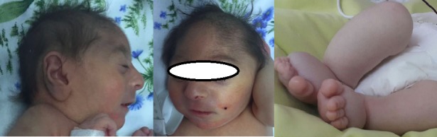
A baby girl was born, on the 35th week of gestation via cesarean section, to an 18-year old mother. Apgar score at the 1st and 5th minute was 8 and 9, respectively. Antenatal scan at 20th gestational week found microcephaly, edema in both lower extremities and the dilation of the proximal intestinal loops. No pathology was found on FISH examination in relation to chromosome 13, 18, 21, X and Y during amniocentesis. At birth baby had weight of 1890 gram (10-50 percentile), the height of 40cm (<10 percentile) and head circumference of 26cm (<10 percentile). Examination of the head revealed microcephaly, micrognathia and a high-bridged nose (Fig. 1). Edema was seen in both lower extremities (Fig. 1). CBC showed thrombocytopenia (86.000/mm3). Liver and kidney function tests, and albumin level were in normal range. Serologic tests for TORCH and Parvovirus were negative. Abdominal ultrasonography (USG) showed bilateral renal hypodysplasia. Ventricular septal defect was found on Echocardiography. Ophthalmologic examination showed microphthalmia, microcornea, and sclerocornea.

 求助内容:
求助内容: 应助结果提醒方式:


