Ling Yu, Mingquan Yan, Jennifer Simkin, Paulina D Ketcham, Eric Leininger, Manjong Han, Ken Muneoka
{"title":"Angiogenesis is inhibitory for mammalian digit regeneration.","authors":"Ling Yu, Mingquan Yan, Jennifer Simkin, Paulina D Ketcham, Eric Leininger, Manjong Han, Ken Muneoka","doi":"10.1002/reg2.24","DOIUrl":null,"url":null,"abstract":"<p><p>The regenerating mouse digit tip is a unique model for investigating blastema formation and epimorphic regeneration in mammals. The blastema is characteristically avascular and we previously reported that blastema expression of a known anti-angiogenic factor gene, Pedf, correlated with a successful regenerative response (Yu, L., Han, M., Yan, M., Lee, E. C., Lee, J. & Muneoka, K. (2010). BMP signaling induces digit regeneration in neonatal mice. Development, 137, 551-559). Here we show that during regeneration Vegfa transcripts are not detected in the blastema but are expressed at the onset of differentiation. Treating the amputation wound with vascular endothelial growth factor enhances angiogenesis but inhibits regeneration. We next tested bone morphogenetic protein 9 (BMP9), another known mediator of angiogenesis, and found that BMP9 is also a potent inhibitor of digit tip regeneration. BMP9 induces Vegfa expression in the digit stump suggesting that regenerative failure is mediated by enhanced angiogenesis. Finally, we show that BMP9 inhibition of regeneration is completely rescued by treatment with pigment epithelium-derived factor. These studies show that precocious angiogenesis is inhibitory for regeneration, and provide compelling evidence that the regulation of angiogenesis is a critical factor in designing therapies aimed at stimulating mammalian regeneration. </p>","PeriodicalId":90316,"journal":{"name":"Regeneration (Oxford, England)","volume":null,"pages":null},"PeriodicalIF":0.0000,"publicationDate":"2014-10-12","publicationTypes":"Journal Article","fieldsOfStudy":null,"isOpenAccess":false,"openAccessPdf":"https://sci-hub-pdf.com/10.1002/reg2.24","citationCount":"35","resultStr":null,"platform":"Semanticscholar","paperid":null,"PeriodicalName":"Regeneration (Oxford, England)","FirstCategoryId":"1085","ListUrlMain":"https://doi.org/10.1002/reg2.24","RegionNum":0,"RegionCategory":null,"ArticlePicture":[],"TitleCN":null,"AbstractTextCN":null,"PMCID":null,"EPubDate":"2014/6/1 0:00:00","PubModel":"eCollection","JCR":"","JCRName":"","Score":null,"Total":0}
引用次数: 35
Abstract
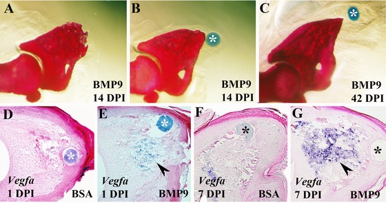
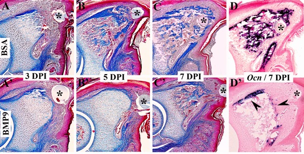
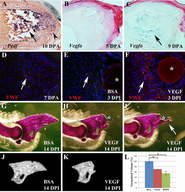
The regenerating mouse digit tip is a unique model for investigating blastema formation and epimorphic regeneration in mammals. The blastema is characteristically avascular and we previously reported that blastema expression of a known anti-angiogenic factor gene, Pedf, correlated with a successful regenerative response (Yu, L., Han, M., Yan, M., Lee, E. C., Lee, J. & Muneoka, K. (2010). BMP signaling induces digit regeneration in neonatal mice. Development, 137, 551-559). Here we show that during regeneration Vegfa transcripts are not detected in the blastema but are expressed at the onset of differentiation. Treating the amputation wound with vascular endothelial growth factor enhances angiogenesis but inhibits regeneration. We next tested bone morphogenetic protein 9 (BMP9), another known mediator of angiogenesis, and found that BMP9 is also a potent inhibitor of digit tip regeneration. BMP9 induces Vegfa expression in the digit stump suggesting that regenerative failure is mediated by enhanced angiogenesis. Finally, we show that BMP9 inhibition of regeneration is completely rescued by treatment with pigment epithelium-derived factor. These studies show that precocious angiogenesis is inhibitory for regeneration, and provide compelling evidence that the regulation of angiogenesis is a critical factor in designing therapies aimed at stimulating mammalian regeneration.

 求助内容:
求助内容: 应助结果提醒方式:


