Juha K Rantala, Sunjong Kwon, James Korkola, Joe W Gray
{"title":"Expanding the Diversity of Imaging-Based RNAi Screen Applications Using Cell Spot Microarrays.","authors":"Juha K Rantala, Sunjong Kwon, James Korkola, Joe W Gray","doi":"10.3390/microarrays2020097","DOIUrl":null,"url":null,"abstract":"<p><p>Over the past decade, great strides have been made in identifying gene aberrations and deregulated pathways that are associated with specific disease states. These association studies guide experimental studies aimed at identifying the aberrant genes and networks that cause the disease states. This requires functional manipulation of these genes and networks in laboratory models of normal and diseased cells. One approach is to assess molecular and biological responses to high-throughput RNA interference (RNAi)-induced gene knockdown. These responses can be revealed by immunofluorescent staining for a molecular or cellular process of interest and quantified using fluorescence image analysis. These applications are typically performed in multiwell format, but are limited by high reagent costs and long plate processing times. These limitations can be mitigated by analyzing cells grown in cell spot microarray (CSMA) format. CSMAs are produced by growing cells on small (~200 mm diameter) spots with each spot carrying an siRNA with transfection reagent. The spacing between spots is only a few hundred micrometers, thus thousands of cell spots can be arranged on a single cell culture surface. These high-density cell cultures can be immunofluorescently stained with minimal reagent consumption and analyzed quickly using automated fluorescence microscopy platforms. This review covers basic aspects of imaging-based CSMA technology, describes a wide range of immunofluorescence assays that have already been implemented successfully for CSMA screening and suggests future directions for advanced RNAi screening experiments. </p>","PeriodicalId":56355,"journal":{"name":"Microarrays","volume":"2 2","pages":"97-114"},"PeriodicalIF":0.0000,"publicationDate":"2013-04-11","publicationTypes":"Journal Article","fieldsOfStudy":null,"isOpenAccess":false,"openAccessPdf":"https://sci-hub-pdf.com/10.3390/microarrays2020097","citationCount":"10","resultStr":null,"platform":"Semanticscholar","paperid":null,"PeriodicalName":"Microarrays","FirstCategoryId":"1085","ListUrlMain":"https://doi.org/10.3390/microarrays2020097","RegionNum":0,"RegionCategory":null,"ArticlePicture":[],"TitleCN":null,"AbstractTextCN":null,"PMCID":null,"EPubDate":"","PubModel":"","JCR":"","JCRName":"","Score":null,"Total":0}
引用次数: 10
Abstract
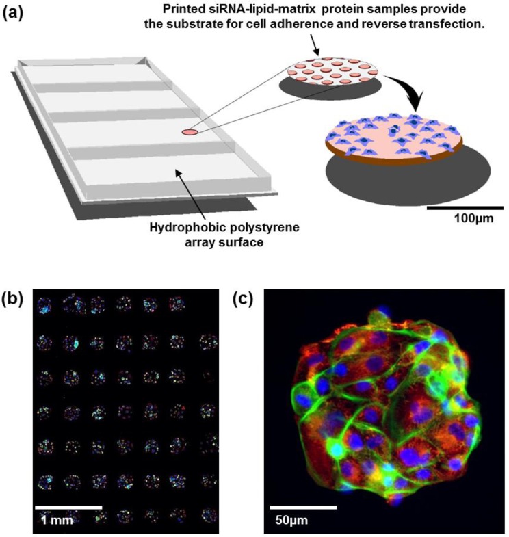
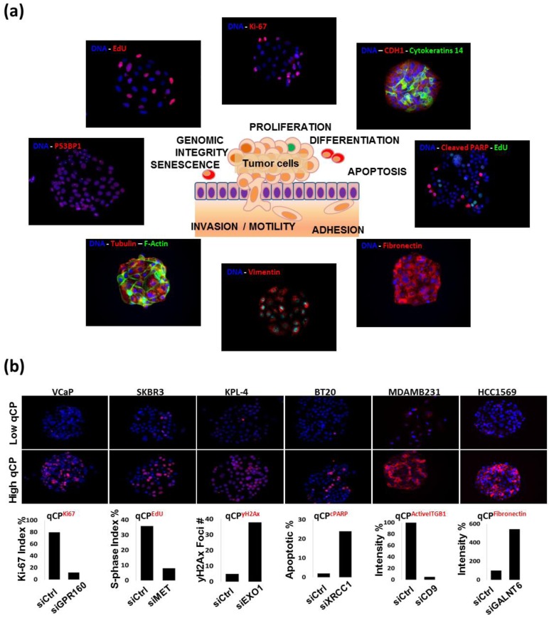
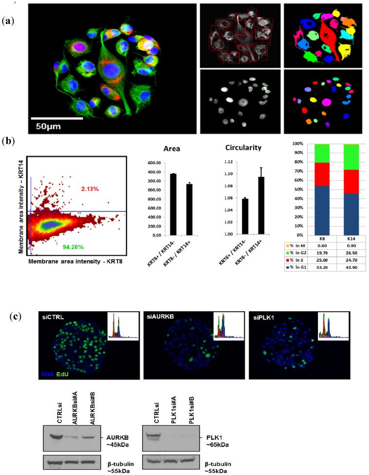
Over the past decade, great strides have been made in identifying gene aberrations and deregulated pathways that are associated with specific disease states. These association studies guide experimental studies aimed at identifying the aberrant genes and networks that cause the disease states. This requires functional manipulation of these genes and networks in laboratory models of normal and diseased cells. One approach is to assess molecular and biological responses to high-throughput RNA interference (RNAi)-induced gene knockdown. These responses can be revealed by immunofluorescent staining for a molecular or cellular process of interest and quantified using fluorescence image analysis. These applications are typically performed in multiwell format, but are limited by high reagent costs and long plate processing times. These limitations can be mitigated by analyzing cells grown in cell spot microarray (CSMA) format. CSMAs are produced by growing cells on small (~200 mm diameter) spots with each spot carrying an siRNA with transfection reagent. The spacing between spots is only a few hundred micrometers, thus thousands of cell spots can be arranged on a single cell culture surface. These high-density cell cultures can be immunofluorescently stained with minimal reagent consumption and analyzed quickly using automated fluorescence microscopy platforms. This review covers basic aspects of imaging-based CSMA technology, describes a wide range of immunofluorescence assays that have already been implemented successfully for CSMA screening and suggests future directions for advanced RNAi screening experiments.
期刊介绍:
High-Throughput (formerly Microarrays, ISSN 2076-3905) is a multidisciplinary peer-reviewed scientific journal that provides an advanced forum for the publication of studies reporting high-dimensional approaches and developments in Life Sciences, Chemistry and related fields. Our aim is to encourage scientists to publish their experimental and theoretical results based on high-throughput techniques as well as computational and statistical tools for data analysis and interpretation. The full experimental or methodological details must be provided so that the results can be reproduced. There is no restriction on the length of the papers. High-Throughput invites submissions covering several topics, including, but not limited to: Microarrays, DNA Sequencing, RNA Sequencing, Protein Identification and Quantification, Cell-based Approaches, Omics Technologies, Imaging, Bioinformatics, Computational Biology/Chemistry, Statistics, Integrative Omics, Drug Discovery and Development, Microfluidics, Lab-on-a-chip, Data Mining, Databases, Multiplex Assays.

 求助内容:
求助内容: 应助结果提醒方式:


