{"title":"Retrieval of mandibular third molar tooth accidentally displaced in submandibular space: Series of two cases.","authors":"Ravinder Solanki, Monika Khangwal, Davender Kumar, Mahesh Goel","doi":"10.4103/0975-962X.184653","DOIUrl":null,"url":null,"abstract":"<p><p>Displacement of tooth or root in submandibular or parapharyngeal spaces is one of the serious complications while extracting mandibular third molar by the general practitioners. Possibilities enhance in cases with extremely thin lingual plates. Moreover, there are no posterior fascial borders limiting the sublingual and submandibular spaces. In addition, no fascial border separates these spaces from the inferior parapharyngeal space. Thus, there is free communication between these spaces and tooth easily may dislodge into further spaces and lead to serious complications ahead. Patients may represent with pain and swelling of the submandibular region and sometimes the limitation in mouth opening when the patient had undergone an unsuccessful surgical procedure and third molar displacement into submandibular space. Two cases of such complications are presented ahead. On clinical examination, submandibular area on the left side of the mandible was tender on palpation. Panoramic radiographs elicited presence of a radiopaque mass similar to that of a tooth root. The computed tomography (CT) scan confirmed the presence of a high-density area in the submandibular region. Orthopantomography and cone beam CT in another patient revealed the displaced third molar in submandibular space. Patients were planned to retrieve the tooth under local anesthesia and the postoperative course was uneventful. </p>","PeriodicalId":90526,"journal":{"name":"Indian journal of dentistry","volume":"7 2","pages":"105-8"},"PeriodicalIF":0.0000,"publicationDate":"2016-04-01","publicationTypes":"Journal Article","fieldsOfStudy":null,"isOpenAccess":false,"openAccessPdf":"https://ftp.ncbi.nlm.nih.gov/pub/pmc/oa_pdf/61/dd/IJDENT-7-105.PMC4934085.pdf","citationCount":"10","resultStr":null,"platform":"Semanticscholar","paperid":null,"PeriodicalName":"Indian journal of dentistry","FirstCategoryId":"1085","ListUrlMain":"https://doi.org/10.4103/0975-962X.184653","RegionNum":0,"RegionCategory":null,"ArticlePicture":[],"TitleCN":null,"AbstractTextCN":null,"PMCID":null,"EPubDate":"","PubModel":"","JCR":"","JCRName":"","Score":null,"Total":0}
引用次数: 10
Abstract
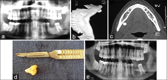
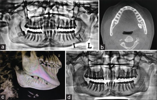
Displacement of tooth or root in submandibular or parapharyngeal spaces is one of the serious complications while extracting mandibular third molar by the general practitioners. Possibilities enhance in cases with extremely thin lingual plates. Moreover, there are no posterior fascial borders limiting the sublingual and submandibular spaces. In addition, no fascial border separates these spaces from the inferior parapharyngeal space. Thus, there is free communication between these spaces and tooth easily may dislodge into further spaces and lead to serious complications ahead. Patients may represent with pain and swelling of the submandibular region and sometimes the limitation in mouth opening when the patient had undergone an unsuccessful surgical procedure and third molar displacement into submandibular space. Two cases of such complications are presented ahead. On clinical examination, submandibular area on the left side of the mandible was tender on palpation. Panoramic radiographs elicited presence of a radiopaque mass similar to that of a tooth root. The computed tomography (CT) scan confirmed the presence of a high-density area in the submandibular region. Orthopantomography and cone beam CT in another patient revealed the displaced third molar in submandibular space. Patients were planned to retrieve the tooth under local anesthesia and the postoperative course was uneventful.

 求助内容:
求助内容: 应助结果提醒方式:


