Sacrococcygeal Teratoma: Mistreated With Repeated Aspirations.
APSP journal of case reports
Pub Date : 2016-06-15
eCollection Date: 2016-07-01
DOI:10.21699/ajcr.v7i3.422
引用次数: 0
Abstract
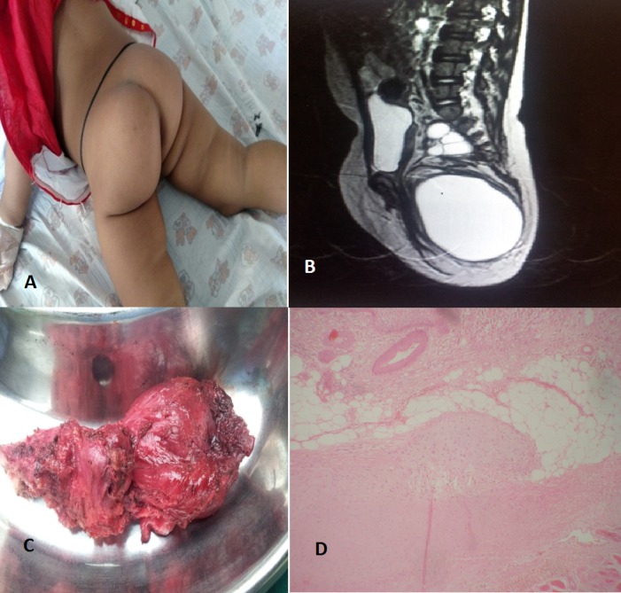
One year and nine month old baby girl brought to the hospital with swelling in the left gluteal region noticed by the parents since three months of age. They consulted a local practitioner who suggested aspiration by needle, which was done multiple times. The size of the swelling used to decrease immediately after aspiration but returned to its usual size within 1-2 months. On examination, the child was alert, active with normal growth and development. Left gluteal region was more prominent with skin over the region showing multiple puncture marks (Fig. 1), palpation revealed an illdefined, immobile, and non-tender mass with variable consistency. Per rectal examination showed mass extending up to the presacral region. Bimanual palpation revealed extension of the mass up to the level of the pelvic brim. Ultrasound revealed a well-defined anechoic cystic lesion near coccyx. MRI of the left gluteal region showed two peripherally enhancing lesions near the anterior-inferior part of the coccyx confirming the diagnosis of SCT (Fig. 1). Serum alpha fetoprotein level was normal. At operation, a large dumb-bell shaped mass with both solid and cystic (mainly) components, arising from the coccyx, was completely excised along with coccygectomy. Postoperative recovery was uneventful. Biopsy report showed it to be mature teratoma (Fig. 1) with margins free of tumor. The parents were counseled for regular follow-up.
骶尾骨畸胎瘤:反复志向的错误治疗。
本文章由计算机程序翻译,如有差异,请以英文原文为准。
求助全文
约1分钟内获得全文
求助全文

 求助内容:
求助内容: 应助结果提醒方式:


