{"title":"Etodolac Containing Topical Niosomal Gel: Formulation Development and Evaluation.","authors":"Gyati Shilakari Asthana, Abhay Asthana, Davinder Singh, Parveen Kumar Sharma","doi":"10.1155/2016/9324567","DOIUrl":null,"url":null,"abstract":"<p><p>The present study aimed to investigate the delivery potential of Etodolac (ETD) containing topical niosomal gel. Niosomal formulations were prepared by thin film hydration method at various ratios of cholesterol and Span 60 and were evaluated with respect to particle size, shape, entrapment efficiency, and in vitro characteristics. Dicetyl phosphate (DCP) was also added in the niosomal formulation. Mean particle size of niosomal formulation was found to be in the range of 2 μm to 4 μm. Niosomal formulation N2 (1 : 1) ratio of cholesterol and surfactant displayed good entrapment efficiency (96.72%). TEM analyses showed that niosomal formulation was spherical in shape. Niosomal formulation (N2) displayed high percentage of drug release after 24 h (94.91) at (1 : 1) ratio of cholesterol : surfactant. Further selected niosomal formulation was used to formulate topical gel and was characterized with respect to its various parameters such as pH, viscosity, spreadability, ex vivo study, and in vivo potential permeation. Ex vivo study showed that niosomal gel possessed better skin permeation study than the plain topical gel. Further in vivo study revealed good inhibition of inflammation in case of topical niosomal gel than plain gel and niosomal formulation. The present study suggested that topical niosomal gel formulations provide sustained and prolonged delivery of drug. </p>","PeriodicalId":15575,"journal":{"name":"Journal of drug delivery","volume":"2016 ","pages":"9324567"},"PeriodicalIF":0.0000,"publicationDate":"2016-01-01","publicationTypes":"Journal Article","fieldsOfStudy":null,"isOpenAccess":false,"openAccessPdf":"https://sci-hub-pdf.com/10.1155/2016/9324567","citationCount":"50","resultStr":null,"platform":"Semanticscholar","paperid":null,"PeriodicalName":"Journal of drug delivery","FirstCategoryId":"1085","ListUrlMain":"https://doi.org/10.1155/2016/9324567","RegionNum":0,"RegionCategory":null,"ArticlePicture":[],"TitleCN":null,"AbstractTextCN":null,"PMCID":null,"EPubDate":"2016/7/11 0:00:00","PubModel":"Epub","JCR":"","JCRName":"","Score":null,"Total":0}
引用次数: 50
Abstract
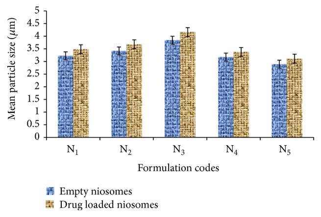
The present study aimed to investigate the delivery potential of Etodolac (ETD) containing topical niosomal gel. Niosomal formulations were prepared by thin film hydration method at various ratios of cholesterol and Span 60 and were evaluated with respect to particle size, shape, entrapment efficiency, and in vitro characteristics. Dicetyl phosphate (DCP) was also added in the niosomal formulation. Mean particle size of niosomal formulation was found to be in the range of 2 μm to 4 μm. Niosomal formulation N2 (1 : 1) ratio of cholesterol and surfactant displayed good entrapment efficiency (96.72%). TEM analyses showed that niosomal formulation was spherical in shape. Niosomal formulation (N2) displayed high percentage of drug release after 24 h (94.91) at (1 : 1) ratio of cholesterol : surfactant. Further selected niosomal formulation was used to formulate topical gel and was characterized with respect to its various parameters such as pH, viscosity, spreadability, ex vivo study, and in vivo potential permeation. Ex vivo study showed that niosomal gel possessed better skin permeation study than the plain topical gel. Further in vivo study revealed good inhibition of inflammation in case of topical niosomal gel than plain gel and niosomal formulation. The present study suggested that topical niosomal gel formulations provide sustained and prolonged delivery of drug.

 求助内容:
求助内容: 应助结果提醒方式:


