Syeda H. Afroze , Ram R. Kalagiri , Michelle Reyes , Jacqueline D. Zimmerman , Madhava R. Beeram , Nathan Drever , David C. Zawieja , Thomas J. Kuehl , Mohammad N. Uddin
{"title":"Apoptotic and stress signaling markers are augmented in preeclamptic placenta and umbilical cord","authors":"Syeda H. Afroze , Ram R. Kalagiri , Michelle Reyes , Jacqueline D. Zimmerman , Madhava R. Beeram , Nathan Drever , David C. Zawieja , Thomas J. Kuehl , Mohammad N. Uddin","doi":"10.1016/j.bbacli.2016.05.003","DOIUrl":null,"url":null,"abstract":"<div><h3>Objective</h3><p>Preeclampsia (preE) has a significant link to alterations of placental function leading to stress and apoptotic signaling, which pass the placental barrier and leave persistent defect in the circulation of the offspring. We assessed apoptotic signaling in placentas and umbilical cords from patients with and without preE.</p></div><div><h3>Methods</h3><p>We collected placental and cord tissues from 27 normal pregnant (NP) women and 20 preE consenting patients after delivery in an IRB approved prospective study. p38 mitogen-activated protein kinase (p38 MAPK) phosphorylation, pro-apoptotic Bcl-2-associated X (Bax), anti-apoptotic Bcl-2, caspase-9, and pro-inflammatory cyclooxygenase-2 (Cox-2) were evaluated by western blot and immunohistochemistry. Comparisons were performed using Student's <em>t</em>-test.</p></div><div><h3>Results</h3><p>p38 phosphorylation (Placenta: 1.5 fold, Cord: 1.7 fold), ratio of Bax/Bcl-2 (Placenta: 1.7 fold, Cord: 2.2 fold), caspase-9 (Placenta: 1.5 fold, Cord: 1.8 fold) and Cox-2 (Placenta: 2.5 fold, Cord: 2.3 fold) were up-regulated (p<!--> <!--><<!--> <!-->0.05) in preE compared to NP patients. Average hospital stays for preE babies were longer than NP babies. No complications were reported for NP babies; however, all of preE babies had multiple complications.</p></div><div><h3>Conclusions</h3><p>Apoptotic and stress signaling are augmented in preE placenta and cord tissue that alter the intrauterine environment and activates the detrimental signaling that is transported to fetus.</p></div>","PeriodicalId":72344,"journal":{"name":"BBA clinical","volume":"6 ","pages":"Pages 25-30"},"PeriodicalIF":0.0000,"publicationDate":"2016-12-01","publicationTypes":"Journal Article","fieldsOfStudy":null,"isOpenAccess":false,"openAccessPdf":"https://sci-hub-pdf.com/10.1016/j.bbacli.2016.05.003","citationCount":"30","resultStr":null,"platform":"Semanticscholar","paperid":null,"PeriodicalName":"BBA clinical","FirstCategoryId":"1085","ListUrlMain":"https://www.sciencedirect.com/science/article/pii/S2214647416300241","RegionNum":0,"RegionCategory":null,"ArticlePicture":[],"TitleCN":null,"AbstractTextCN":null,"PMCID":null,"EPubDate":"","PubModel":"","JCR":"","JCRName":"","Score":null,"Total":0}
引用次数: 30
Abstract
Objective
Preeclampsia (preE) has a significant link to alterations of placental function leading to stress and apoptotic signaling, which pass the placental barrier and leave persistent defect in the circulation of the offspring. We assessed apoptotic signaling in placentas and umbilical cords from patients with and without preE.
Methods
We collected placental and cord tissues from 27 normal pregnant (NP) women and 20 preE consenting patients after delivery in an IRB approved prospective study. p38 mitogen-activated protein kinase (p38 MAPK) phosphorylation, pro-apoptotic Bcl-2-associated X (Bax), anti-apoptotic Bcl-2, caspase-9, and pro-inflammatory cyclooxygenase-2 (Cox-2) were evaluated by western blot and immunohistochemistry. Comparisons were performed using Student's t-test.
Results
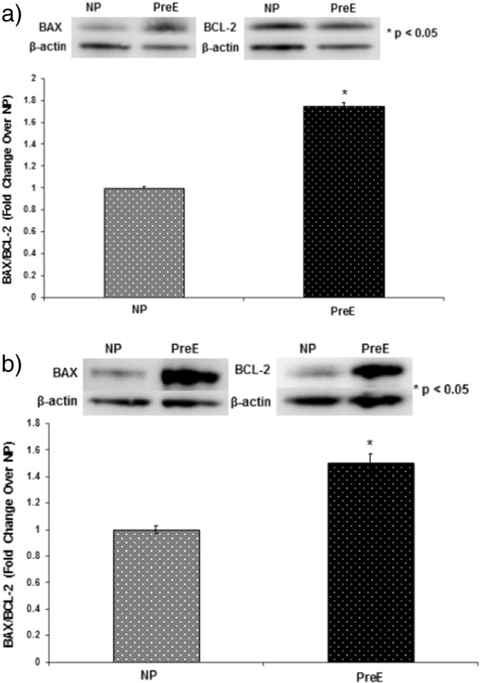
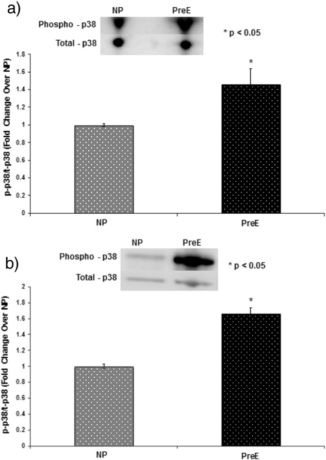
p38 phosphorylation (Placenta: 1.5 fold, Cord: 1.7 fold), ratio of Bax/Bcl-2 (Placenta: 1.7 fold, Cord: 2.2 fold), caspase-9 (Placenta: 1.5 fold, Cord: 1.8 fold) and Cox-2 (Placenta: 2.5 fold, Cord: 2.3 fold) were up-regulated (p < 0.05) in preE compared to NP patients. Average hospital stays for preE babies were longer than NP babies. No complications were reported for NP babies; however, all of preE babies had multiple complications.
Conclusions
Apoptotic and stress signaling are augmented in preE placenta and cord tissue that alter the intrauterine environment and activates the detrimental signaling that is transported to fetus.

 求助内容:
求助内容: 应助结果提醒方式:


