Jae Hyun Kwon, Shin Hwang, Gi-Won Song, Deok-Bog Moon, Gil-Chun Park, Seok-Hwan Kim, Sung-Gyu Lee
{"title":"Conjoined unification venoplasty for triple portal vein branches of right liver graft: a case report and technical refinement.","authors":"Jae Hyun Kwon, Shin Hwang, Gi-Won Song, Deok-Bog Moon, Gil-Chun Park, Seok-Hwan Kim, Sung-Gyu Lee","doi":"10.14701/kjhbps.2016.20.2.61","DOIUrl":null,"url":null,"abstract":"<p><p>Anomalous portal vein (PV) branching of the donor liver is uncommon and usually makes two, or rarely, more separate PV branches at the right liver graft. Autologous PV Y-graft interposition has long been regarded as the standard procedure, but is currently replaced with the newly developed technique of conjoined unification venoplasty (CUV) due to its superior results. Herein, we presented a case of CUV application to three PV openings of a right liver graft. The recipient was a 32-year-old male patient with hepatitis B virus-associated liver cirrhosis. The living liver donor was his 33-year-old sister who had a type III PV anomaly, but the right posterior PV branch was bifurcated early into separate branches of the segments VI and VII, thus three right liver PV branches were cut separately. We used the CUV technique consisting of placement of a small vein unification patch between three PV orifices, followed by overlying coverage with a crotch-opened autologous portal Y-graft. The portal Y-graft was excised and its crotches were incised to make a wide common orifice. Three bidirectional running sutures were required to attach the crotch-opened autologous portal Y-graft. After portal reperfusion, the conjoined PV portion bulged like a tennis ball, providing a wide range of alignment tolerance. The patient recovered uneventfully from the liver transplantation operation. The CUV technique enabled uneventful reconstruction of triple donor PV orifices. Thus, CUV can be a useful and effective technical option for reconstruction of right liver grafts with various anomalous PVs. </p>","PeriodicalId":91136,"journal":{"name":"Korean journal of hepato-biliary-pancreatic surgery","volume":"20 2","pages":"61-5"},"PeriodicalIF":0.0000,"publicationDate":"2016-05-01","publicationTypes":"Journal Article","fieldsOfStudy":null,"isOpenAccess":false,"openAccessPdf":"https://sci-hub-pdf.com/10.14701/kjhbps.2016.20.2.61","citationCount":"6","resultStr":null,"platform":"Semanticscholar","paperid":null,"PeriodicalName":"Korean journal of hepato-biliary-pancreatic surgery","FirstCategoryId":"1085","ListUrlMain":"https://doi.org/10.14701/kjhbps.2016.20.2.61","RegionNum":0,"RegionCategory":null,"ArticlePicture":[],"TitleCN":null,"AbstractTextCN":null,"PMCID":null,"EPubDate":"2016/5/11 0:00:00","PubModel":"Epub","JCR":"","JCRName":"","Score":null,"Total":0}
引用次数: 6
Abstract
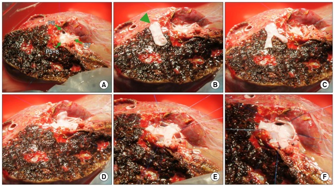
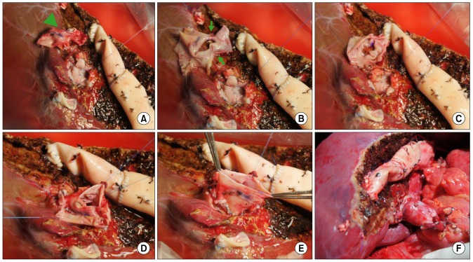
Anomalous portal vein (PV) branching of the donor liver is uncommon and usually makes two, or rarely, more separate PV branches at the right liver graft. Autologous PV Y-graft interposition has long been regarded as the standard procedure, but is currently replaced with the newly developed technique of conjoined unification venoplasty (CUV) due to its superior results. Herein, we presented a case of CUV application to three PV openings of a right liver graft. The recipient was a 32-year-old male patient with hepatitis B virus-associated liver cirrhosis. The living liver donor was his 33-year-old sister who had a type III PV anomaly, but the right posterior PV branch was bifurcated early into separate branches of the segments VI and VII, thus three right liver PV branches were cut separately. We used the CUV technique consisting of placement of a small vein unification patch between three PV orifices, followed by overlying coverage with a crotch-opened autologous portal Y-graft. The portal Y-graft was excised and its crotches were incised to make a wide common orifice. Three bidirectional running sutures were required to attach the crotch-opened autologous portal Y-graft. After portal reperfusion, the conjoined PV portion bulged like a tennis ball, providing a wide range of alignment tolerance. The patient recovered uneventfully from the liver transplantation operation. The CUV technique enabled uneventful reconstruction of triple donor PV orifices. Thus, CUV can be a useful and effective technical option for reconstruction of right liver grafts with various anomalous PVs.


 求助内容:
求助内容: 应助结果提醒方式:


