Luis Miguel Sousa Marques, Gonçalo Neto d'Almeida, José Cabral
{"title":"\"Two-step\" technique with OsiriX™ to evaluate feasibility of C2 pedicle for surgical fixation.","authors":"Luis Miguel Sousa Marques, Gonçalo Neto d'Almeida, José Cabral","doi":"10.4103/0974-8237.181826","DOIUrl":null,"url":null,"abstract":"<p><strong>Background: </strong>Surgical treatment of craniovertebral junction pathology has evolved considerably in recent decades with the implementation of short atlanto-axial fixation techniques, notwhithstanding increasing neurovascular risks. Also, there is strong evidence that fixation of C2 anatomical pedicle has the best biomechanical profile of the entire cervical spine. However, it is often difficult and misleading, to evaluate anatomical bony and vascular anomalies using the three orthogonal planes (axial, coronal, and sagittal) of CT.</p><p><strong>Objectives: </strong>The authors describe an innovative and simple technique to evaluate the feasibility of C2 pedicle for surgical screw fixation using preoperative planning with the free DICOM (Digital Imaging and Communications in Medicine) software OsiriX™.</p><p><strong>Materials and methods: </strong>The authors report the applicatin of this novel technique in 5 cases (3 traumatic, 1 Os Odontoideum, and 1 complex congenital malformation) collected from our general case series of the Department in the last 5 years.</p><p><strong>Results: </strong>In this proof of concept study, the pre-operative analysis with the two-step tecnique was detrimental for choosing the surgical tecnique. Detailed post-operative analysis confirmed correct position of C2 screws without cortical breach. There were no complications or mortality reported.</p><p><strong>Conclusion: </strong>This two-step technique is an easy and reliable way to determine the feasibility of C2 pedicle for surgical fixation. The detailed tridimensional radiological preoperative evaluation of craniovertebral junction anatomy is critical to the sucess and safety of this surgeries, and can avoid, to certain degree, expensive intra-operative tridimensional imaging facilities.</p>","PeriodicalId":520667,"journal":{"name":"Journal of craniovertebral junction & spine","volume":" ","pages":"75-81"},"PeriodicalIF":1.3000,"publicationDate":"2016-04-01","publicationTypes":"Journal Article","fieldsOfStudy":null,"isOpenAccess":false,"openAccessPdf":"https://ftp.ncbi.nlm.nih.gov/pub/pmc/oa_pdf/42/96/JCVJS-7-75.PMC4872566.pdf","citationCount":"7","resultStr":null,"platform":"Semanticscholar","paperid":null,"PeriodicalName":"Journal of craniovertebral junction & spine","FirstCategoryId":"1085","ListUrlMain":"https://doi.org/10.4103/0974-8237.181826","RegionNum":0,"RegionCategory":null,"ArticlePicture":[],"TitleCN":null,"AbstractTextCN":null,"PMCID":null,"EPubDate":"","PubModel":"","JCR":"","JCRName":"","Score":null,"Total":0}
引用次数: 7
Abstract
Background: Surgical treatment of craniovertebral junction pathology has evolved considerably in recent decades with the implementation of short atlanto-axial fixation techniques, notwhithstanding increasing neurovascular risks. Also, there is strong evidence that fixation of C2 anatomical pedicle has the best biomechanical profile of the entire cervical spine. However, it is often difficult and misleading, to evaluate anatomical bony and vascular anomalies using the three orthogonal planes (axial, coronal, and sagittal) of CT.
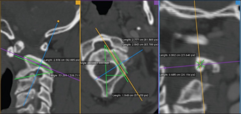
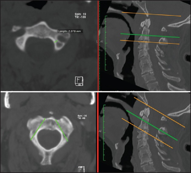
Objectives: The authors describe an innovative and simple technique to evaluate the feasibility of C2 pedicle for surgical screw fixation using preoperative planning with the free DICOM (Digital Imaging and Communications in Medicine) software OsiriX™.
Materials and methods: The authors report the applicatin of this novel technique in 5 cases (3 traumatic, 1 Os Odontoideum, and 1 complex congenital malformation) collected from our general case series of the Department in the last 5 years.
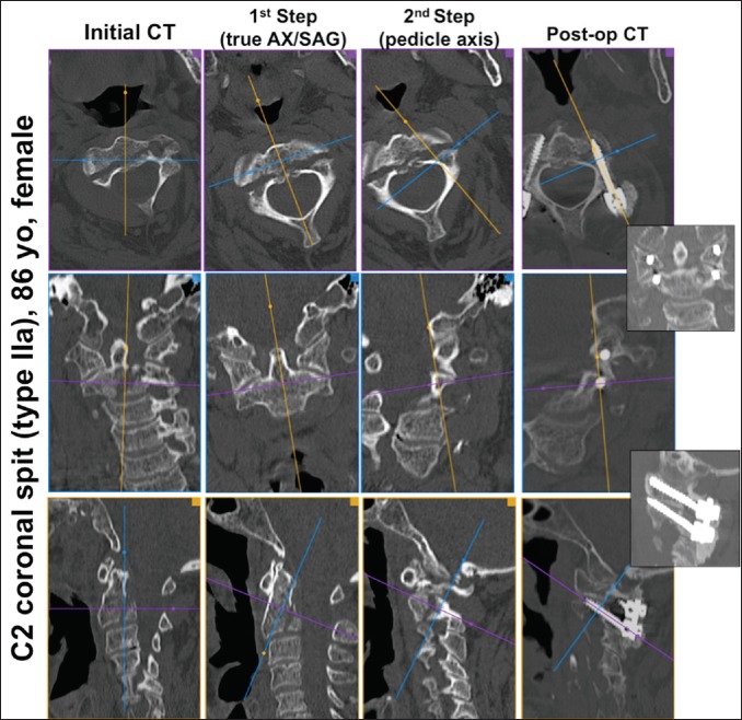
Results: In this proof of concept study, the pre-operative analysis with the two-step tecnique was detrimental for choosing the surgical tecnique. Detailed post-operative analysis confirmed correct position of C2 screws without cortical breach. There were no complications or mortality reported.
Conclusion: This two-step technique is an easy and reliable way to determine the feasibility of C2 pedicle for surgical fixation. The detailed tridimensional radiological preoperative evaluation of craniovertebral junction anatomy is critical to the sucess and safety of this surgeries, and can avoid, to certain degree, expensive intra-operative tridimensional imaging facilities.

 求助内容:
求助内容: 应助结果提醒方式:


