Immunohistochemical expression of vascular endothelial growth factor in keratocystic odontogenic tumor, dentigerous cyst, and radicular cyst: A comparative study.
{"title":"Immunohistochemical expression of vascular endothelial growth factor in keratocystic odontogenic tumor, dentigerous cyst, and radicular cyst: A comparative study.","authors":"Nidhi Khajuria, Rashmi Metgud, Smitha Naik, Sahul Lerra, Priya Tiwari, Mamta, Payal Katakwar, Anirudh Tak","doi":"10.4103/0975-962X.179378","DOIUrl":null,"url":null,"abstract":"<p><strong>Background: </strong>Cyst and tumors arise from tissue remains of odontogenesis, these interactions have been considered to play an important role in the tumorigenesis of odontogenic lesions. The connective tissue stroma has an essential role in the preservation of epithelial tissues and minor alterations in the epithelium are followed by corresponding changes in the stroma, such as angiogenesis. Vascular endothelial growth factor (VEGF) is considered the first factor which maintains its position as the most critical driver of vascular formation and is required to initiate the formation of immature vessels, with this aim, present study was executed to evaluate VEGF expression in kertocystic odontogenic tumor, dentigerous cyst and radicular cyst (RC).</p><p><strong>Materials and methods: </strong>A retrospective study was carried out comprising a total of 31 cases; 13 cases of keratocystic odontogenic tumor (KCOT), nine cases of dentigerous cyst (DC) and nine cases of RC. The sections were stained immunohistochemically with VEGF antibody and were evaluated for the presence and intensity of the immuno reactive cells. Statistical analysis was carried out using Chi-square test to inter-compare the VEGF expression between KCOT, DC, and RC.</p><p><strong>Results: </strong>VEGF expression in the epithelium and connective tissue was significantly higher in KCOT compared to dentigerous and RC. One case of KCOT with carcinomatous change also revealed positive results for the VEGF expression in the dysplastic epithelium, tumor islands, and connective tissue. The significant difference was observed on inter-comparison of the VEGF expression in the connective tissue of KCOT and DC, whereas no significant difference was observed in the VEGF expression in the connective tissue of KCOT and DC.</p><p><strong>Conclusion: </strong>The present study data supports the literature finding that angiogenesis can be important in the progression and enlargement of odontogenic cysts similarly to what occurs in neoplastic conditions and further it can be concluded that the higher positivity for VEGF of KCOT could help to explain in part the aggressive biological behavior of the lesion. The stroma of KCOT could be regarded not only as a structural support of the cyst wall but also as playing a part in the neoplastic behavior of cyst.</p>","PeriodicalId":90526,"journal":{"name":"Indian journal of dentistry","volume":"7 1","pages":"17-22"},"PeriodicalIF":0.0000,"publicationDate":"2016-01-01","publicationTypes":"Journal Article","fieldsOfStudy":null,"isOpenAccess":false,"openAccessPdf":"https://ftp.ncbi.nlm.nih.gov/pub/pmc/oa_pdf/47/05/IJDENT-7-17.PMC4836093.pdf","citationCount":"3","resultStr":null,"platform":"Semanticscholar","paperid":null,"PeriodicalName":"Indian journal of dentistry","FirstCategoryId":"1085","ListUrlMain":"https://doi.org/10.4103/0975-962X.179378","RegionNum":0,"RegionCategory":null,"ArticlePicture":[],"TitleCN":null,"AbstractTextCN":null,"PMCID":null,"EPubDate":"","PubModel":"","JCR":"","JCRName":"","Score":null,"Total":0}
引用次数: 3
Abstract
Background: Cyst and tumors arise from tissue remains of odontogenesis, these interactions have been considered to play an important role in the tumorigenesis of odontogenic lesions. The connective tissue stroma has an essential role in the preservation of epithelial tissues and minor alterations in the epithelium are followed by corresponding changes in the stroma, such as angiogenesis. Vascular endothelial growth factor (VEGF) is considered the first factor which maintains its position as the most critical driver of vascular formation and is required to initiate the formation of immature vessels, with this aim, present study was executed to evaluate VEGF expression in kertocystic odontogenic tumor, dentigerous cyst and radicular cyst (RC).
Materials and methods: A retrospective study was carried out comprising a total of 31 cases; 13 cases of keratocystic odontogenic tumor (KCOT), nine cases of dentigerous cyst (DC) and nine cases of RC. The sections were stained immunohistochemically with VEGF antibody and were evaluated for the presence and intensity of the immuno reactive cells. Statistical analysis was carried out using Chi-square test to inter-compare the VEGF expression between KCOT, DC, and RC.
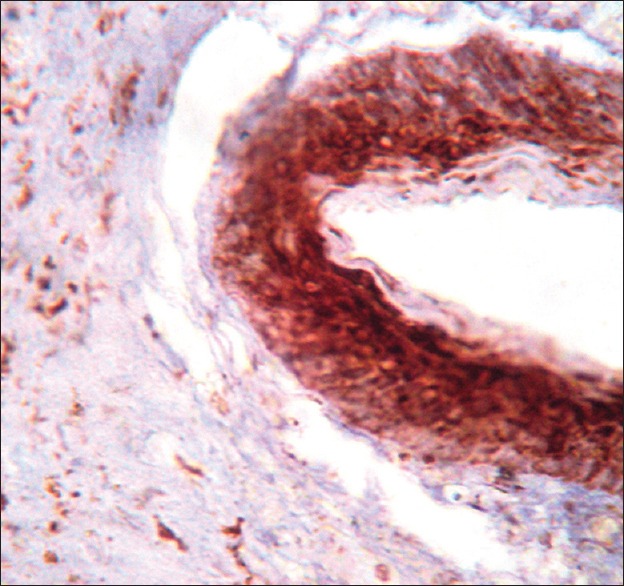
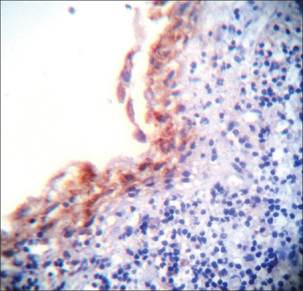
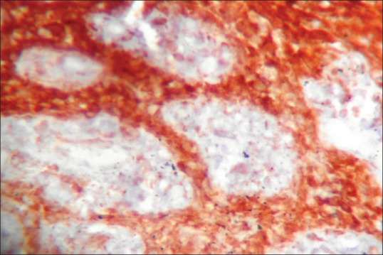
Results: VEGF expression in the epithelium and connective tissue was significantly higher in KCOT compared to dentigerous and RC. One case of KCOT with carcinomatous change also revealed positive results for the VEGF expression in the dysplastic epithelium, tumor islands, and connective tissue. The significant difference was observed on inter-comparison of the VEGF expression in the connective tissue of KCOT and DC, whereas no significant difference was observed in the VEGF expression in the connective tissue of KCOT and DC.
Conclusion: The present study data supports the literature finding that angiogenesis can be important in the progression and enlargement of odontogenic cysts similarly to what occurs in neoplastic conditions and further it can be concluded that the higher positivity for VEGF of KCOT could help to explain in part the aggressive biological behavior of the lesion. The stroma of KCOT could be regarded not only as a structural support of the cyst wall but also as playing a part in the neoplastic behavior of cyst.

 求助内容:
求助内容: 应助结果提醒方式:


