Hongmei Li-Byarlay, Barry R Pittendrigh, Larry L Murdock
{"title":"Plant Defense Inhibitors Affect the Structures of Midgut Cells in Drosophila melanogaster and Callosobruchus maculatus.","authors":"Hongmei Li-Byarlay, Barry R Pittendrigh, Larry L Murdock","doi":"10.4137/IJIS.S28595","DOIUrl":null,"url":null,"abstract":"<p><p>Plants produce proteins such as protease inhibitors and lectins as defenses against herbivorous insects and pathogens. However, no systematic studies have explored the structural responses in the midguts of insects when challenged with plant defensive proteins and lectins across different species. In this study, we fed two kinds of protease inhibitors and lectins to the fruit fly Drosophila melanogaster and alpha-amylase inhibitors and lectins to the cowpea bruchid Callosobruchus maculatus. We assessed the changes in midgut cell structures by comparing them with such structures in insects receiving normal diets or subjected to food deprivation. Using light and transmission electron microscopy in both species, we observed structural changes in the midgut peritrophic matrix as well as shortened microvilli on the surfaces of midgut epithelial cells in D. melanogaster. Dietary inhibitors and lectins caused similar lesions in the epithelial cells but not much change in the peritrophic matrix in both species. We also noted structural damages in the Drosophila midgut after six hours of starvation and changes were still present after 12 hours. Our study provided the first evidence of key structural changes of midguts using a comparative approach between a dipteran and a coleopteran. Our particular observation and discussion on plant-insect interaction and dietary stress are relevant for future mode of action studies of plant defensive protein in insect physiology. </p>","PeriodicalId":73456,"journal":{"name":"International journal of insect science","volume":"8 ","pages":"71-9"},"PeriodicalIF":0.0000,"publicationDate":"2016-08-29","publicationTypes":"Journal Article","fieldsOfStudy":null,"isOpenAccess":false,"openAccessPdf":"https://sci-hub-pdf.com/10.4137/IJIS.S28595","citationCount":"7","resultStr":null,"platform":"Semanticscholar","paperid":null,"PeriodicalName":"International journal of insect science","FirstCategoryId":"1085","ListUrlMain":"https://doi.org/10.4137/IJIS.S28595","RegionNum":0,"RegionCategory":null,"ArticlePicture":[],"TitleCN":null,"AbstractTextCN":null,"PMCID":null,"EPubDate":"2016/1/1 0:00:00","PubModel":"eCollection","JCR":"","JCRName":"","Score":null,"Total":0}
引用次数: 7
Abstract
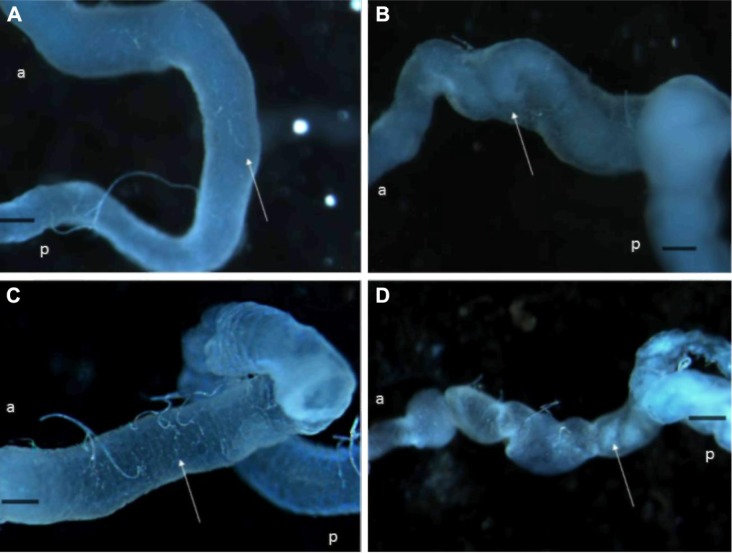
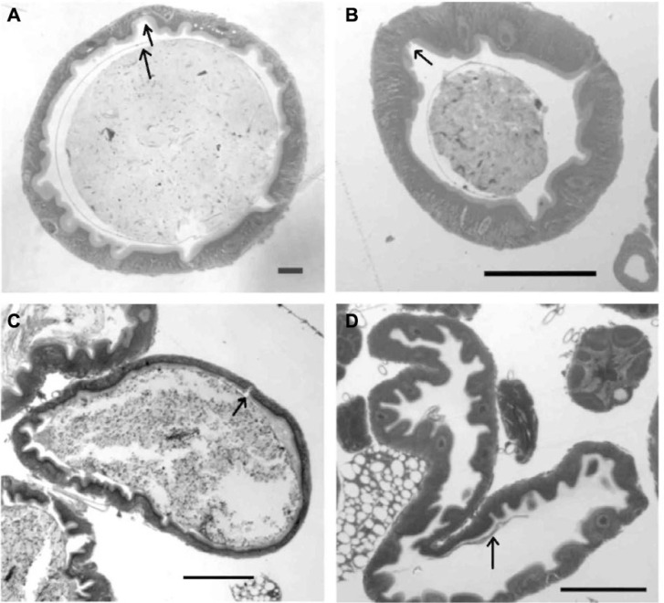
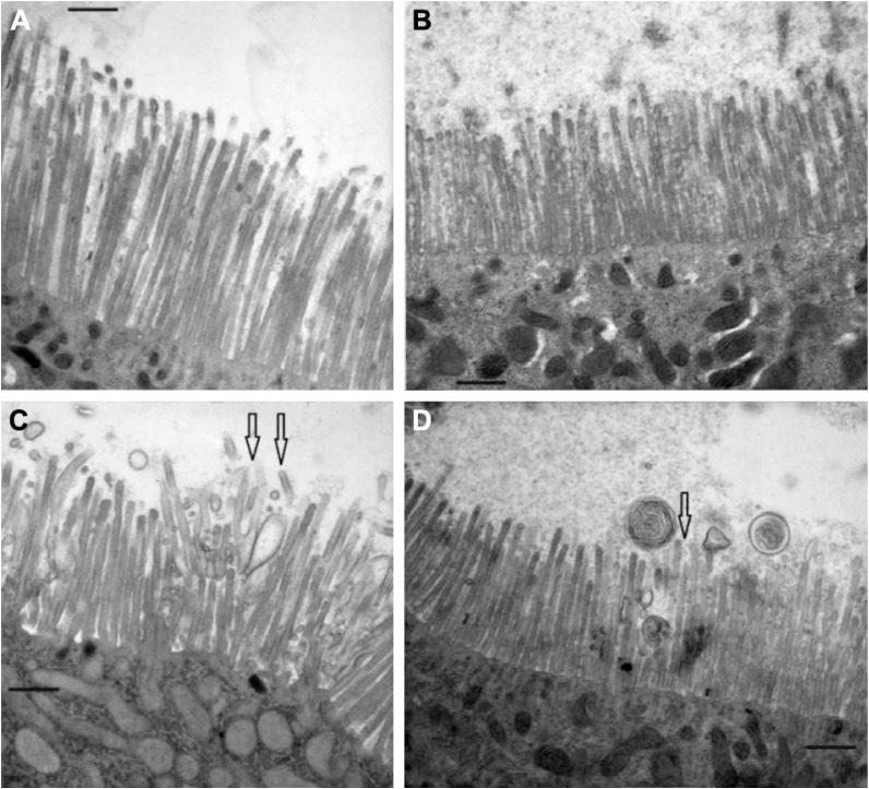
Plants produce proteins such as protease inhibitors and lectins as defenses against herbivorous insects and pathogens. However, no systematic studies have explored the structural responses in the midguts of insects when challenged with plant defensive proteins and lectins across different species. In this study, we fed two kinds of protease inhibitors and lectins to the fruit fly Drosophila melanogaster and alpha-amylase inhibitors and lectins to the cowpea bruchid Callosobruchus maculatus. We assessed the changes in midgut cell structures by comparing them with such structures in insects receiving normal diets or subjected to food deprivation. Using light and transmission electron microscopy in both species, we observed structural changes in the midgut peritrophic matrix as well as shortened microvilli on the surfaces of midgut epithelial cells in D. melanogaster. Dietary inhibitors and lectins caused similar lesions in the epithelial cells but not much change in the peritrophic matrix in both species. We also noted structural damages in the Drosophila midgut after six hours of starvation and changes were still present after 12 hours. Our study provided the first evidence of key structural changes of midguts using a comparative approach between a dipteran and a coleopteran. Our particular observation and discussion on plant-insect interaction and dietary stress are relevant for future mode of action studies of plant defensive protein in insect physiology.

 求助内容:
求助内容: 应助结果提醒方式:


