Dysregulation of Protein Kinase Gene Expression in NK Cells from Chronic Fatigue Syndrome/Myalgic Encephalomyelitis Patients.
Gene regulation and systems biology
Pub Date : 2016-08-28
eCollection Date: 2016-01-01
DOI:10.4137/GRSB.S40036
引用次数: 11
Abstract
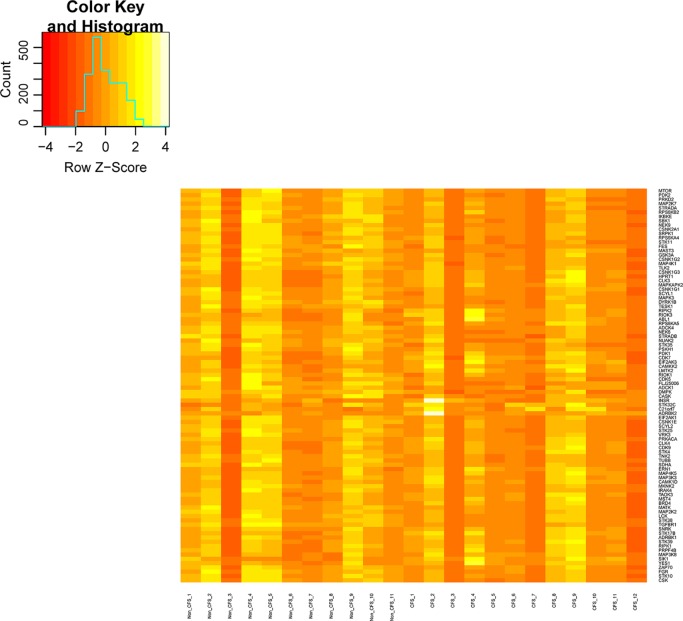
BACKGROUND The etiology and pathomechanism of chronic fatigue syndrome/myalgic encephalomyelitis (CFS/ME) are unknown. However, natural killer (NK) cell dysfunction, in particular reduced NK cytotoxic activity, is a consistent finding in CFS/ME patients. Previous research has reported significant changes in intracellular mitogen-activated protein kinase pathways from isolated NK cells. The purpose of this present investigation was to examine whether protein kinase genes have a role in abnormal NK cell intracellular signaling in CFS/ME. METHOD Messenger RNA (mRNA) expression of 528 protein kinase genes in isolated NK cells was analyzed (nCounter GX Human Kinase Kit v2 (XT); NanoString Technologies) from moderate (n = 11; age, 54.9 ± 10.3 years) and severe (n = 12; age, 47.5 ± 8.0 years) CFS/ME patients (classified by the 2011 International Consensus Criteria) and nonfatigued controls (n = 11; age, 50.0 ± 12.3 years). RESULTS The expression of 92 protein kinase genes was significantly different in the severe CFS/ME group compared with nonfatigued controls. Among these, 37 genes were significantly upregulated and 55 genes were significantly downregulated in severe CFS/ME patients compared with nonfatigued controls. CONCLUSIONS In severe CFS/ME patients, dysfunction in protein kinase genes may contribute to impairments in NK cell intracellular signaling and effector function. Similar changes in protein kinase genes may be present in other cells, potentially contributing to the pathomechanism of this illness.

慢性疲劳综合征/肌痛性脑脊髓炎患者NK细胞蛋白激酶基因表达失调
背景:慢性疲劳综合征/肌痛性脑脊髓炎(CFS/ME)的病因和病理机制尚不清楚。然而,自然杀伤(NK)细胞功能障碍,特别是NK细胞毒性活性降低,在CFS/ME患者中是一致的发现。先前的研究报道了分离NK细胞的细胞内有丝分裂原激活的蛋白激酶途径的显著变化。本研究的目的是研究蛋白激酶基因是否在CFS/ME中异常NK细胞胞内信号传导中起作用。方法:采用nCounter GX Human kinase Kit v2 (XT)检测分离NK细胞中528个蛋白激酶基因的mRNA表达;NanoString Technologies) from moderate (n = 11;年龄(54.9±10.3岁)和重度(n = 12;年龄(47.5±8.0岁)CFS/ME患者(按2011年国际共识标准分类)和非疲劳对照组(n = 11;年龄:50.0±12.3岁。结果:与非疲劳对照组相比,重度CFS/ME组92个蛋白激酶基因的表达有显著差异。其中,与非疲劳对照相比,重度CFS/ME患者中有37个基因显著上调,55个基因显著下调。结论:在严重CFS/ME患者中,蛋白激酶基因功能障碍可能导致NK细胞胞内信号通路和效应物功能受损。蛋白激酶基因的类似变化可能存在于其他细胞中,可能有助于这种疾病的病理机制。
本文章由计算机程序翻译,如有差异,请以英文原文为准。
求助全文
约1分钟内获得全文
求助全文

 求助内容:
求助内容: 应助结果提醒方式:


