{"title":"Malignant Transformation of Vagal Nerve Schwannoma in to Angiosarcoma: A Rare Event.","authors":"Sangeet Kumar Agarwal, Manish Munjal, Devinder Rai, Seema Rao","doi":"10.4103/2006-8808.184941","DOIUrl":null,"url":null,"abstract":"<p><p>Schwannomas are benign, rare peripheral nerve sheath tumors that occur in the head and neck region. Some physicians opt to closely observe cases of schwannoma of the neck on an outpatient basis rather than to perform radical surgery. However, there is a possibility, albeit rare, of malignant transformation of a benign schwannoma. Here, we are reporting the first case from the Indian subcontinent which was transformed into the angiosarcoma from benign vagal schwannoma over a long period. A 47-year-old male patient complaining of left sided neck swelling since last 12 years, swelling was insidious in onset, gradually progressive very slowly. In last 2 months, the size of the swelling was suddenly increased. On examination, there was an approximately 6 cm × 6 cm of size, firm, nodular, well-defined, nontender swelling in the left lateral part of the neck. Fine-needle aspiration cytology (FNAC) revealed paraganglioma and magnetic resonance imaging demonstrated very clearly a tumor, its morphology, and its relation to the surrounding structures, the tumor was thought to be a vagal schwannoma. Surgery was done, and the whole of the tumor was removed in toto. On final histopathological diagnosis, the tumor was proved to be angiosarcoma developed from vagal schwannoma. Postoperative chemotherapy was given but due to distant metastasis, the patient died. Long standing neck masses can convert into malignancy as in our case, therefore, work up of the patient should be done properly. Multiple FNAC should be done because single FNAC can give the false negative result as in our case. This was our diagnostic drawback not to do multiple computed tomography guided FNAC. </p>","PeriodicalId":89430,"journal":{"name":"Journal of surgical technique and case report","volume":"7 1","pages":"17-9"},"PeriodicalIF":0.0000,"publicationDate":"2015-01-01","publicationTypes":"Journal Article","fieldsOfStudy":null,"isOpenAccess":false,"openAccessPdf":"https://ftp.ncbi.nlm.nih.gov/pub/pmc/oa_pdf/7f/a1/JSTCR-7-17.PMC4959406.pdf","citationCount":"5","resultStr":null,"platform":"Semanticscholar","paperid":null,"PeriodicalName":"Journal of surgical technique and case report","FirstCategoryId":"1085","ListUrlMain":"https://doi.org/10.4103/2006-8808.184941","RegionNum":0,"RegionCategory":null,"ArticlePicture":[],"TitleCN":null,"AbstractTextCN":null,"PMCID":null,"EPubDate":"","PubModel":"","JCR":"","JCRName":"","Score":null,"Total":0}
引用次数: 5
Abstract
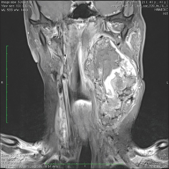
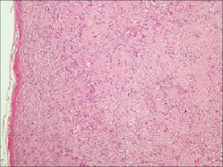
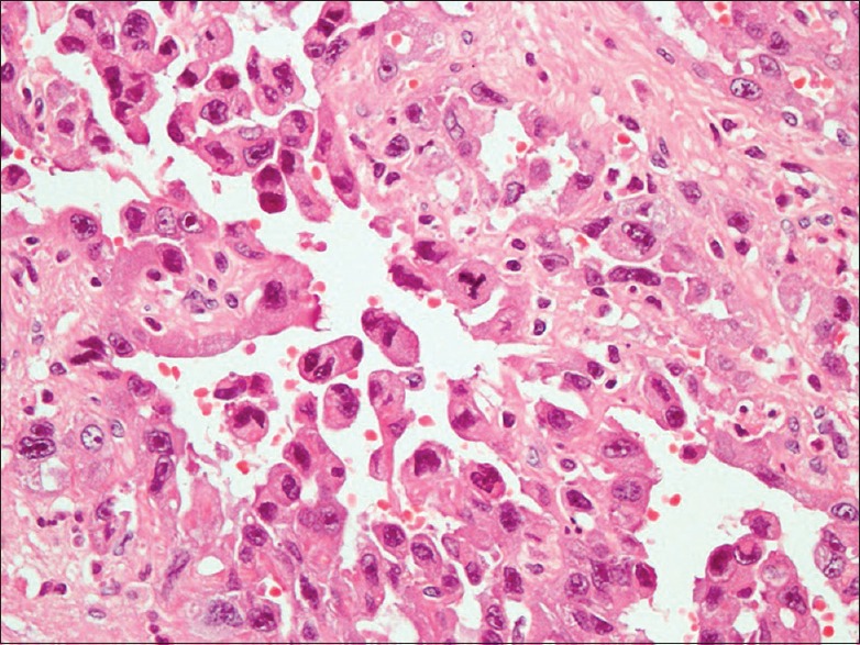
Schwannomas are benign, rare peripheral nerve sheath tumors that occur in the head and neck region. Some physicians opt to closely observe cases of schwannoma of the neck on an outpatient basis rather than to perform radical surgery. However, there is a possibility, albeit rare, of malignant transformation of a benign schwannoma. Here, we are reporting the first case from the Indian subcontinent which was transformed into the angiosarcoma from benign vagal schwannoma over a long period. A 47-year-old male patient complaining of left sided neck swelling since last 12 years, swelling was insidious in onset, gradually progressive very slowly. In last 2 months, the size of the swelling was suddenly increased. On examination, there was an approximately 6 cm × 6 cm of size, firm, nodular, well-defined, nontender swelling in the left lateral part of the neck. Fine-needle aspiration cytology (FNAC) revealed paraganglioma and magnetic resonance imaging demonstrated very clearly a tumor, its morphology, and its relation to the surrounding structures, the tumor was thought to be a vagal schwannoma. Surgery was done, and the whole of the tumor was removed in toto. On final histopathological diagnosis, the tumor was proved to be angiosarcoma developed from vagal schwannoma. Postoperative chemotherapy was given but due to distant metastasis, the patient died. Long standing neck masses can convert into malignancy as in our case, therefore, work up of the patient should be done properly. Multiple FNAC should be done because single FNAC can give the false negative result as in our case. This was our diagnostic drawback not to do multiple computed tomography guided FNAC.

 求助内容:
求助内容: 应助结果提醒方式:


