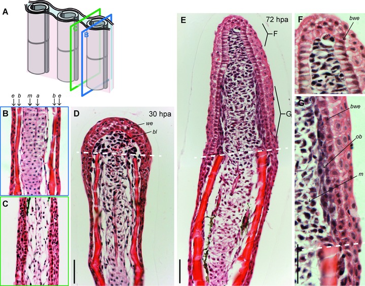
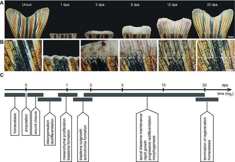
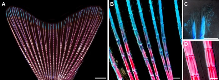
{"title":"The art of fin regeneration in zebrafish.","authors":"Catherine Pfefferli, Anna Jaźwińska","doi":"10.1002/reg2.33","DOIUrl":null,"url":null,"abstract":"<p><p>The zebrafish fin provides a valuable model to study the epimorphic type of regeneration, whereby the amputated part of the appendage is nearly perfectly replaced. To accomplish fin regeneration, two reciprocally interacting domains need to be established at the injury site, namely a wound epithelium and a blastema. The wound epithelium provides a supporting niche for the blastema, which contains mesenchyme-derived progenitor cells for the regenerate. The fate of blastemal daughter cells depends on their relative position with respect to the fin margin. The apical compartment of the outgrowth maintains its undifferentiated character, whereas the proximal descendants of the blastema progressively switch from the proliferation program to the morphogenesis program. A delicate balance between self-renewal and differentiation has to be continuously adjusted during the course of regeneration. This review summarizes the current knowledge about the cellular and molecular mechanisms of blastema formation, and discusses several studies related to the regulation of growth and morphogenesis during fin regeneration. A wide range of canonical signaling pathways has been implicated during the establishment and maintenance of the blastema. Epigenetic mechanisms play a crucial role in the regulation of cellular plasticity during the transition between differentiation states. Ion fluxes, gap-junctional communication and protein phosphatase activity have been shown to coordinate proliferation and tissue patterning in the caudal fin. The identification of the downstream targets of the fin regeneration signals and the discovery of mechanisms integrating the variety of input pathways represent exciting future aims in this fascinating field of research. </p>","PeriodicalId":90316,"journal":{"name":"Regeneration (Oxford, England)","volume":"2 2","pages":"72-83"},"PeriodicalIF":0.0000,"publicationDate":"2015-05-19","publicationTypes":"Journal Article","fieldsOfStudy":null,"isOpenAccess":false,"openAccessPdf":"https://sci-hub-pdf.com/10.1002/reg2.33","citationCount":"175","resultStr":null,"platform":"Semanticscholar","paperid":null,"PeriodicalName":"Regeneration (Oxford, England)","FirstCategoryId":"1085","ListUrlMain":"https://doi.org/10.1002/reg2.33","RegionNum":0,"RegionCategory":null,"ArticlePicture":[],"TitleCN":null,"AbstractTextCN":null,"PMCID":null,"EPubDate":"2015/4/1 0:00:00","PubModel":"eCollection","JCR":"","JCRName":"","Score":null,"Total":0}
引用次数: 175
Abstract
The zebrafish fin provides a valuable model to study the epimorphic type of regeneration, whereby the amputated part of the appendage is nearly perfectly replaced. To accomplish fin regeneration, two reciprocally interacting domains need to be established at the injury site, namely a wound epithelium and a blastema. The wound epithelium provides a supporting niche for the blastema, which contains mesenchyme-derived progenitor cells for the regenerate. The fate of blastemal daughter cells depends on their relative position with respect to the fin margin. The apical compartment of the outgrowth maintains its undifferentiated character, whereas the proximal descendants of the blastema progressively switch from the proliferation program to the morphogenesis program. A delicate balance between self-renewal and differentiation has to be continuously adjusted during the course of regeneration. This review summarizes the current knowledge about the cellular and molecular mechanisms of blastema formation, and discusses several studies related to the regulation of growth and morphogenesis during fin regeneration. A wide range of canonical signaling pathways has been implicated during the establishment and maintenance of the blastema. Epigenetic mechanisms play a crucial role in the regulation of cellular plasticity during the transition between differentiation states. Ion fluxes, gap-junctional communication and protein phosphatase activity have been shown to coordinate proliferation and tissue patterning in the caudal fin. The identification of the downstream targets of the fin regeneration signals and the discovery of mechanisms integrating the variety of input pathways represent exciting future aims in this fascinating field of research.

 求助内容:
求助内容: 应助结果提醒方式:


