Posterior Spinal Reconstruction with Pedicle Screws, Multiple Iliac Screws and Wisconsin Spinal Wires in a Patient with Neurofibromatosis Scoliosis: A Case Report.
Woong-Beom Kim, Young-Seop Park, Jong-Hwa Park, Seung-Jae Hyun
{"title":"Posterior Spinal Reconstruction with Pedicle Screws, Multiple Iliac Screws and Wisconsin Spinal Wires in a Patient with Neurofibromatosis Scoliosis: A Case Report.","authors":"Woong-Beom Kim, Young-Seop Park, Jong-Hwa Park, Seung-Jae Hyun","doi":"10.14245/kjs.2015.12.3.181","DOIUrl":null,"url":null,"abstract":"<p><p>A 54-year-old female with neurofibromatosis type 1 presented with progressing truncal shift owing to spinal deformity. On plain radiograph, the Cobb angle was 54 degree in coronal plane. Radiological examinations showed severe dystrophic change with dysplastic pedicles, bony scalloping, neural foraminal widening from dural ectasia. The patient underwent deformity correction and reconstruction surgery from the T9 to the pelvis using multiple iliac screws and Wisconsin interspinous segmental instrumentation by wiring due to maximize fixation points. The postoperative course was uneventful. One-year follow-up radiographs showed a successful curve correction with solid fusion. We report a case of pedicle dysplasia and dystrophic change treated by posterior segmental spinal instrumentation and fusion with help of multiple iliac screws and modified Wisconsin interspinous segmental wiring. </p>","PeriodicalId":17867,"journal":{"name":"Korean Journal of Spine","volume":"12 3","pages":"181-4"},"PeriodicalIF":0.0000,"publicationDate":"2015-09-01","publicationTypes":"Journal Article","fieldsOfStudy":null,"isOpenAccess":false,"openAccessPdf":"https://ftp.ncbi.nlm.nih.gov/pub/pmc/oa_pdf/c5/1f/kjs-12-181.PMC4623179.pdf","citationCount":"3","resultStr":null,"platform":"Semanticscholar","paperid":null,"PeriodicalName":"Korean Journal of Spine","FirstCategoryId":"1085","ListUrlMain":"https://doi.org/10.14245/kjs.2015.12.3.181","RegionNum":0,"RegionCategory":null,"ArticlePicture":[],"TitleCN":null,"AbstractTextCN":null,"PMCID":null,"EPubDate":"2015/9/30 0:00:00","PubModel":"Epub","JCR":"","JCRName":"","Score":null,"Total":0}
引用次数: 3
Abstract
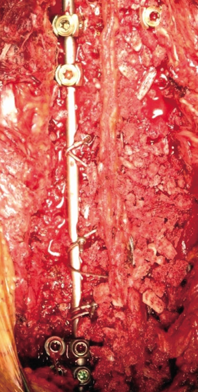
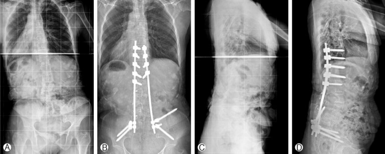
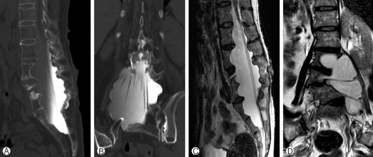
A 54-year-old female with neurofibromatosis type 1 presented with progressing truncal shift owing to spinal deformity. On plain radiograph, the Cobb angle was 54 degree in coronal plane. Radiological examinations showed severe dystrophic change with dysplastic pedicles, bony scalloping, neural foraminal widening from dural ectasia. The patient underwent deformity correction and reconstruction surgery from the T9 to the pelvis using multiple iliac screws and Wisconsin interspinous segmental instrumentation by wiring due to maximize fixation points. The postoperative course was uneventful. One-year follow-up radiographs showed a successful curve correction with solid fusion. We report a case of pedicle dysplasia and dystrophic change treated by posterior segmental spinal instrumentation and fusion with help of multiple iliac screws and modified Wisconsin interspinous segmental wiring.

 求助内容:
求助内容: 应助结果提醒方式:


