Brigitte Bisaro, Giorgia Mandili, Alice Poli, Andrea Piolatto, Valentina Papa, Francesco Novelli, Giovanna Cenacchi, Marco Forni, Cristina Zanini
{"title":"Proteomic analysis of extracellular vesicles from medullospheres reveals a role for iron in the cancer progression of medulloblastoma.","authors":"Brigitte Bisaro, Giorgia Mandili, Alice Poli, Andrea Piolatto, Valentina Papa, Francesco Novelli, Giovanna Cenacchi, Marco Forni, Cristina Zanini","doi":"","DOIUrl":null,"url":null,"abstract":"<p><strong>Background: </strong>Medulloblastoma (MB) is the most common malignant childhood brain tumor with the propensity to disseminate at an early stage, and is associated with high morbidity. New treatment strategies are needed to improve cure rates and to reduce life-long cognitive and functional deficits associated with current therapies. Extracellular Vesicles (EVs) are important players in cell-to-cell communication in health and diseases. A clearer understanding of cell-to-cell communication in tumors can be achieved by studying EV secretion in medullospheres. This can reveal subtle modifications induced by the passage from adherent to non-adherent growth, as spheres may account for the adaptation of tumor cells to the mutated environment.</p><p><strong>Methods: </strong>Formation of medullospheres from MB cell lines stabilized in adherent conditions was obtained through culture conditioning based on low attachment flasks and specialized medium. EVs collected by ultracentrifugation, in adherent conditions and as spheres, were subjected to electron microscopy, NanoSight measurements and proteomics.</p><p><strong>Results: </strong>Interestingly, iron carrier proteins were only found in EVs shed by CSC-enriched tumor cell population of spheres. We used iron chelators when culturing MB cell lines as spheres. Iron chelators induced a decrease in number/size of spheres and in stem cell populations able to initiate in vitro spheres formation.</p><p><strong>Conclusions: </strong>This work suggests a not yet identified role of iron metabolism in MB progression and invasion and opens the possibility to use chelators as adjuvants in anti-tumoral chemotherapy.</p>","PeriodicalId":90271,"journal":{"name":"Molecular and cellular therapies","volume":"3 ","pages":"8"},"PeriodicalIF":0.0000,"publicationDate":"2015-10-13","publicationTypes":"Journal Article","fieldsOfStudy":null,"isOpenAccess":false,"openAccessPdf":"https://www.ncbi.nlm.nih.gov/pmc/articles/PMC4603768/pdf/","citationCount":"0","resultStr":null,"platform":"Semanticscholar","paperid":null,"PeriodicalName":"Molecular and cellular therapies","FirstCategoryId":"1085","ListUrlMain":"","RegionNum":0,"RegionCategory":null,"ArticlePicture":[],"TitleCN":null,"AbstractTextCN":null,"PMCID":null,"EPubDate":"2015/1/1 0:00:00","PubModel":"eCollection","JCR":"","JCRName":"","Score":null,"Total":0}
引用次数: 0
Abstract
Background: Medulloblastoma (MB) is the most common malignant childhood brain tumor with the propensity to disseminate at an early stage, and is associated with high morbidity. New treatment strategies are needed to improve cure rates and to reduce life-long cognitive and functional deficits associated with current therapies. Extracellular Vesicles (EVs) are important players in cell-to-cell communication in health and diseases. A clearer understanding of cell-to-cell communication in tumors can be achieved by studying EV secretion in medullospheres. This can reveal subtle modifications induced by the passage from adherent to non-adherent growth, as spheres may account for the adaptation of tumor cells to the mutated environment.
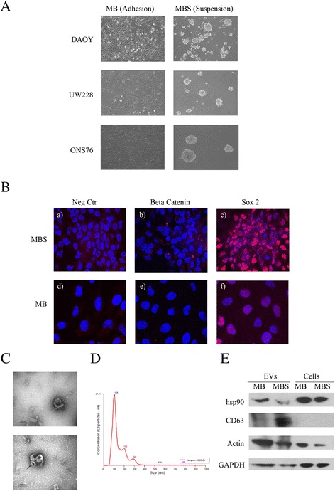
Methods: Formation of medullospheres from MB cell lines stabilized in adherent conditions was obtained through culture conditioning based on low attachment flasks and specialized medium. EVs collected by ultracentrifugation, in adherent conditions and as spheres, were subjected to electron microscopy, NanoSight measurements and proteomics.
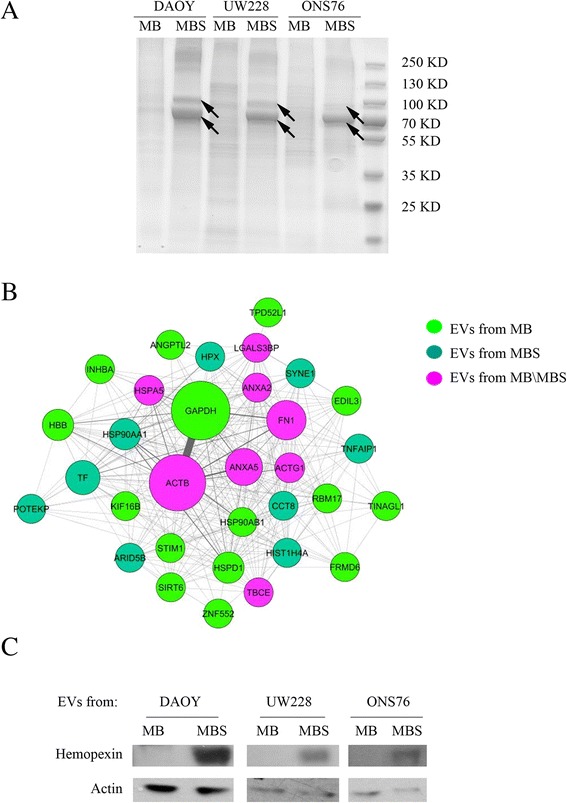
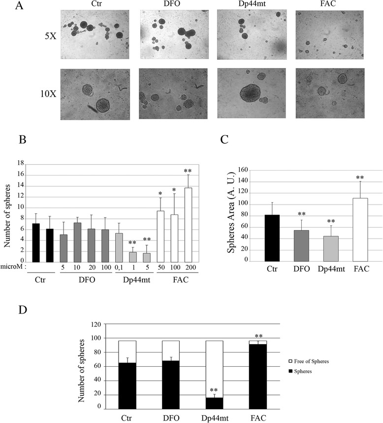
Results: Interestingly, iron carrier proteins were only found in EVs shed by CSC-enriched tumor cell population of spheres. We used iron chelators when culturing MB cell lines as spheres. Iron chelators induced a decrease in number/size of spheres and in stem cell populations able to initiate in vitro spheres formation.
Conclusions: This work suggests a not yet identified role of iron metabolism in MB progression and invasion and opens the possibility to use chelators as adjuvants in anti-tumoral chemotherapy.

 求助内容:
求助内容: 应助结果提醒方式:


