Mesut Sipahi, Sevinç Şahin, Ergin Arslan, Hasan Börekci, Bayram Metin, Nuh Zafer Cantürk
{"title":"Effect of the Human Amniotic Membrane on Liver Regeneration in Rats.","authors":"Mesut Sipahi, Sevinç Şahin, Ergin Arslan, Hasan Börekci, Bayram Metin, Nuh Zafer Cantürk","doi":"10.1155/2015/706186","DOIUrl":null,"url":null,"abstract":"<p><p>Introduction. Operations are performed for broader liver surgery indications for a better understanding of hepatic anatomy/physiology and developments in operation technology. Surgery can cure some patients with liver metastasis of some tumors. Nevertheless, postoperative liver failure is the most feared complication causing mortality in patients who have undergone excision of a large liver mass. The human amniotic membrane has regenerative effects. Thus, we investigated the effects of the human amniotic membrane on regeneration of the resected liver. Methods. Twenty female Wistar albino rats were divided into control and experimental groups and underwent a 70% hepatectomy. The human amniotic membrane was placed over the residual liver in the experimental group. Relative liver weight, histopathological features, and biochemical parameters were assessed on postoperative day 3. Results. Total protein and albumin levels were significantly lower in the experimental group than in the control group. No difference in relative liver weight was observed between the groups. Hepatocyte mitotic count was significantly higher in the experimental group than in the control group. Hepatic steatosis was detected in the experimental group. Conclusion. Applying the amniotic membrane to residual liver adversely affected liver regeneration. However, mesenchymal stem cell research has the potential to accelerate liver regeneration investigations. </p>","PeriodicalId":77165,"journal":{"name":"HPB surgery : a world journal of hepatic, pancreatic and biliary surgery","volume":"2015 ","pages":"706186"},"PeriodicalIF":0.0000,"publicationDate":"2015-01-01","publicationTypes":"Journal Article","fieldsOfStudy":null,"isOpenAccess":false,"openAccessPdf":"https://sci-hub-pdf.com/10.1155/2015/706186","citationCount":"8","resultStr":null,"platform":"Semanticscholar","paperid":null,"PeriodicalName":"HPB surgery : a world journal of hepatic, pancreatic and biliary surgery","FirstCategoryId":"1085","ListUrlMain":"https://doi.org/10.1155/2015/706186","RegionNum":0,"RegionCategory":null,"ArticlePicture":[],"TitleCN":null,"AbstractTextCN":null,"PMCID":null,"EPubDate":"2015/9/17 0:00:00","PubModel":"Epub","JCR":"","JCRName":"","Score":null,"Total":0}
引用次数: 8
Abstract
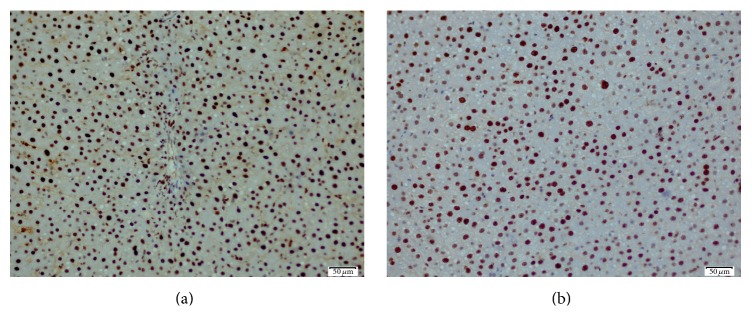
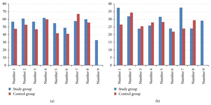
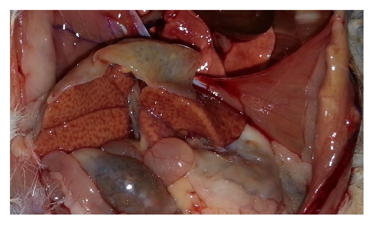
Introduction. Operations are performed for broader liver surgery indications for a better understanding of hepatic anatomy/physiology and developments in operation technology. Surgery can cure some patients with liver metastasis of some tumors. Nevertheless, postoperative liver failure is the most feared complication causing mortality in patients who have undergone excision of a large liver mass. The human amniotic membrane has regenerative effects. Thus, we investigated the effects of the human amniotic membrane on regeneration of the resected liver. Methods. Twenty female Wistar albino rats were divided into control and experimental groups and underwent a 70% hepatectomy. The human amniotic membrane was placed over the residual liver in the experimental group. Relative liver weight, histopathological features, and biochemical parameters were assessed on postoperative day 3. Results. Total protein and albumin levels were significantly lower in the experimental group than in the control group. No difference in relative liver weight was observed between the groups. Hepatocyte mitotic count was significantly higher in the experimental group than in the control group. Hepatic steatosis was detected in the experimental group. Conclusion. Applying the amniotic membrane to residual liver adversely affected liver regeneration. However, mesenchymal stem cell research has the potential to accelerate liver regeneration investigations.

 求助内容:
求助内容: 应助结果提醒方式:


