Velia Ramírez-Amador, Gabriela Anaya-Saavedra, Brenda Crabtree-Ramírez, Lilly Esquivel-Pedraza, Marcela Saeb-Lima, Juan Sierra-Madero
{"title":"Clinical Spectrum of Oral Secondary Syphilis in HIV-Infected Patients.","authors":"Velia Ramírez-Amador, Gabriela Anaya-Saavedra, Brenda Crabtree-Ramírez, Lilly Esquivel-Pedraza, Marcela Saeb-Lima, Juan Sierra-Madero","doi":"10.1155/2013/892427","DOIUrl":null,"url":null,"abstract":"<p><p>Background. Oral lesions may constitute the first clinical manifestation in secondary syphilis, but detailed descriptions in HIV-infected individuals are scarce. Objective. To describe the clinical characteristics of oral secondary syphilis in HIV-infected patients and its relevance in the early diagnosis of syphilis. Methods. Twenty HIV/AIDS adult subjects with oral secondary syphilis lesions presenting at two HIV/AIDS referral centers in Mexico City (2003-2011) are described. An oral examination was performed by specialists in oral pathology and medicine; when possible, a punch biopsy was done, and Warthin-Starry stain and immunohistochemistry were completed. Intraoral herpes virus infection and erythematous candidosis were ruled out by cytological analysis. Diagnosis of oral syphilis was confirmed with positive nontreponemal test (VDRL), and, if possible, fluorescent treponemal antibody test. Results. Twenty male patients (median age 31.5, 21-59 years) with oral secondary syphilis lesions were included. Oral lesions were the first clinical sign of syphilis in 16 (80%) cases. Mucous patch was the most common oral manifestation (17, 85.5%), followed by shallow ulcers (2, 10%) and macular lesions (1, 5%). Conclusions. Due to the recent rise in HIV-syphilis coinfection, dental and medical practitioners should consider secondary syphilis in the differential diagnosis of oral lesions, particularly in HIV-infected patients. </p>","PeriodicalId":90237,"journal":{"name":"Journal of sexually transmitted diseases","volume":"2013 ","pages":"892427"},"PeriodicalIF":0.0000,"publicationDate":"2013-01-01","publicationTypes":"Journal Article","fieldsOfStudy":null,"isOpenAccess":false,"openAccessPdf":"https://sci-hub-pdf.com/10.1155/2013/892427","citationCount":"21","resultStr":null,"platform":"Semanticscholar","paperid":null,"PeriodicalName":"Journal of sexually transmitted diseases","FirstCategoryId":"1085","ListUrlMain":"https://doi.org/10.1155/2013/892427","RegionNum":0,"RegionCategory":null,"ArticlePicture":[],"TitleCN":null,"AbstractTextCN":null,"PMCID":null,"EPubDate":"2012/12/17 0:00:00","PubModel":"Epub","JCR":"","JCRName":"","Score":null,"Total":0}
引用次数: 21
Abstract
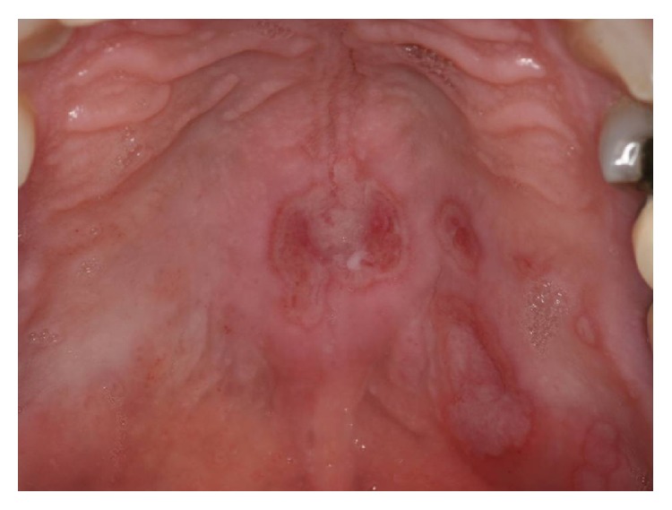
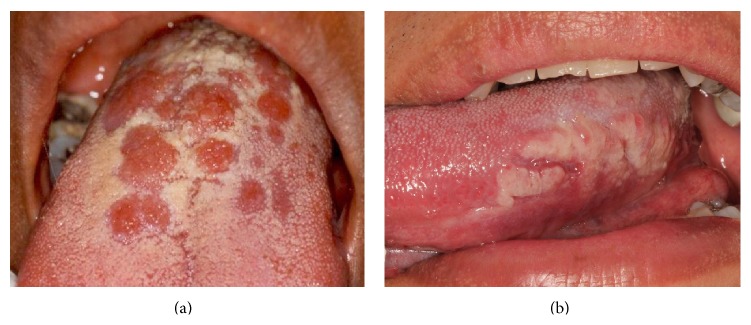
Background. Oral lesions may constitute the first clinical manifestation in secondary syphilis, but detailed descriptions in HIV-infected individuals are scarce. Objective. To describe the clinical characteristics of oral secondary syphilis in HIV-infected patients and its relevance in the early diagnosis of syphilis. Methods. Twenty HIV/AIDS adult subjects with oral secondary syphilis lesions presenting at two HIV/AIDS referral centers in Mexico City (2003-2011) are described. An oral examination was performed by specialists in oral pathology and medicine; when possible, a punch biopsy was done, and Warthin-Starry stain and immunohistochemistry were completed. Intraoral herpes virus infection and erythematous candidosis were ruled out by cytological analysis. Diagnosis of oral syphilis was confirmed with positive nontreponemal test (VDRL), and, if possible, fluorescent treponemal antibody test. Results. Twenty male patients (median age 31.5, 21-59 years) with oral secondary syphilis lesions were included. Oral lesions were the first clinical sign of syphilis in 16 (80%) cases. Mucous patch was the most common oral manifestation (17, 85.5%), followed by shallow ulcers (2, 10%) and macular lesions (1, 5%). Conclusions. Due to the recent rise in HIV-syphilis coinfection, dental and medical practitioners should consider secondary syphilis in the differential diagnosis of oral lesions, particularly in HIV-infected patients.


 求助内容:
求助内容: 应助结果提醒方式:


