{"title":"Differential Diagnosis of Cystic Pancreatic Lesions Including the Usefulness of Biomarkers.","authors":"Philippe Lévy, Vinciane Rebours","doi":"10.1159/000371786","DOIUrl":null,"url":null,"abstract":"<p><strong>Background: </strong>Cystic pancreatic lesions are more and more often found. Malignant risk ranges from nil to more than 60%. A precise diagnosis is required to adapt surveillance or therapeutic strategy.</p><p><strong>Methods: </strong>We tried to identify the most difficult differential diagnoses encountered in a tertiary center of pancreatology and to guide the reader as how to reach the correct strategy and diagnosis in these situations.</p><p><strong>Results: </strong>We identified eight clinically difficult situations: i) chronic pancreatitis versus intraductal papillary mucinous neoplasms, ii) serous versus mucinous cystic neoplasms, iii) serous cystic neoplasms versus branch-duct intraductal papillary mucinous neoplasms, iv) intraductal papillary mucinous neoplasms versus acinar cell cystadenoma, v) (pseudo-) solid serous cystic neoplasm versus neuroendocrine tumor, vi) pancreatic neuroendocrine tumors versus solid pseudopapillary tumors, vii) cystic forms of a solid tumor, and viii) rare pancreatic or peripancreatic cystic lesions. The work-up should rely on computed tomography scan, pancreatic magnetic resonance imaging, and, only if necessary, endoscopic ultrasound with or without fine needle aspiration.</p><p><strong>Conclusion: </strong>An expert analysis of imaging data allows a precise diagnosis in most of the cases. Pancreatic resection should no longer be performed in case of diagnostic doubt.</p>","PeriodicalId":49114,"journal":{"name":"Viszeralmedizin","volume":"31 1","pages":"7-13"},"PeriodicalIF":0.0000,"publicationDate":"2015-02-01","publicationTypes":"Journal Article","fieldsOfStudy":null,"isOpenAccess":false,"openAccessPdf":"https://sci-hub-pdf.com/10.1159/000371786","citationCount":"5","resultStr":null,"platform":"Semanticscholar","paperid":null,"PeriodicalName":"Viszeralmedizin","FirstCategoryId":"1085","ListUrlMain":"https://doi.org/10.1159/000371786","RegionNum":0,"RegionCategory":null,"ArticlePicture":[],"TitleCN":null,"AbstractTextCN":null,"PMCID":null,"EPubDate":"","PubModel":"","JCR":"","JCRName":"","Score":null,"Total":0}
引用次数: 5
Abstract
Background: Cystic pancreatic lesions are more and more often found. Malignant risk ranges from nil to more than 60%. A precise diagnosis is required to adapt surveillance or therapeutic strategy.
Methods: We tried to identify the most difficult differential diagnoses encountered in a tertiary center of pancreatology and to guide the reader as how to reach the correct strategy and diagnosis in these situations.
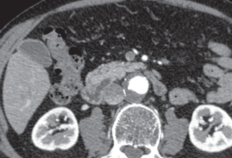
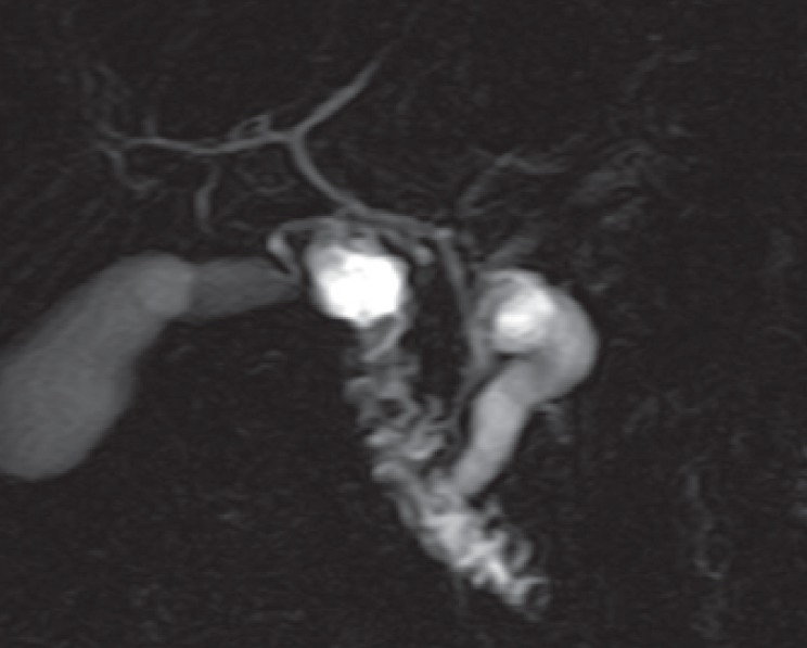
Results: We identified eight clinically difficult situations: i) chronic pancreatitis versus intraductal papillary mucinous neoplasms, ii) serous versus mucinous cystic neoplasms, iii) serous cystic neoplasms versus branch-duct intraductal papillary mucinous neoplasms, iv) intraductal papillary mucinous neoplasms versus acinar cell cystadenoma, v) (pseudo-) solid serous cystic neoplasm versus neuroendocrine tumor, vi) pancreatic neuroendocrine tumors versus solid pseudopapillary tumors, vii) cystic forms of a solid tumor, and viii) rare pancreatic or peripancreatic cystic lesions. The work-up should rely on computed tomography scan, pancreatic magnetic resonance imaging, and, only if necessary, endoscopic ultrasound with or without fine needle aspiration.
Conclusion: An expert analysis of imaging data allows a precise diagnosis in most of the cases. Pancreatic resection should no longer be performed in case of diagnostic doubt.
 求助内容:
求助内容: 应助结果提醒方式:


