Seohyun Kim, Seung Joon Choi, Su Joa Ahn, So Hyun Park, Young Sup Shim, Jeong Ho Kim
{"title":"Clinical Course of Small Subepithelial Tumors of the Small Bowel Detected on CT.","authors":"Seohyun Kim, Seung Joon Choi, Su Joa Ahn, So Hyun Park, Young Sup Shim, Jeong Ho Kim","doi":"10.3348/jksr.2021.0048","DOIUrl":null,"url":null,"abstract":"<p><strong>Purpose: </strong>This study aimed to evaluate the natural growth of subepithelial tumors of the small bowel detected on CT.</p><p><strong>Materials and methods: </strong>Consecutive patients who were suspected of having subepithelial tumors of the small bowel between January 2005 and December 2020 were reviewed. Eligible patients with suspected small (< 30 mm) subepithelial tumors on at least two CT evaluations were included in the analysis. The patients' data on demographic characteristics, tumoral characteristics, and tumoral size changes during the follow-up were collected.</p><p><strong>Results: </strong>This study included 64 patients with suspected small subepithelial tumors (<i>n</i> = 64) of the small bowel. After a median follow-up of 15.8 months, the diameter and volume growth rates were 0.02 mm/month and 1.5 mm<sup>3</sup>/month, respectively. A significant correlation was observed between the initial size and the growth rate of the small bowel subepithelial tumors. The group of large-sized tumors (initial diameter ≥ 10 mm) tended to show lobulated contours, heterogeneous enhancement, and necrotic changes more frequently than the group of small-sized tumors (initial diameter < 10 mm).</p><p><strong>Conclusion: </strong>Small bowel subepithelial tumors measuring less than 10 mm grew more slowly than those measuring 10-30 mm.</p>","PeriodicalId":74904,"journal":{"name":"Taehan Yongsang Uihakhoe chi","volume":" ","pages":"608-619"},"PeriodicalIF":0.0000,"publicationDate":"2022-05-01","publicationTypes":"Journal Article","fieldsOfStudy":null,"isOpenAccess":false,"openAccessPdf":"https://ftp.ncbi.nlm.nih.gov/pub/pmc/oa_pdf/04/41/jksr-83-608.PMC9514538.pdf","citationCount":"0","resultStr":null,"platform":"Semanticscholar","paperid":null,"PeriodicalName":"Taehan Yongsang Uihakhoe chi","FirstCategoryId":"1085","ListUrlMain":"https://doi.org/10.3348/jksr.2021.0048","RegionNum":0,"RegionCategory":null,"ArticlePicture":[],"TitleCN":null,"AbstractTextCN":null,"PMCID":null,"EPubDate":"2021/12/23 0:00:00","PubModel":"Epub","JCR":"","JCRName":"","Score":null,"Total":0}
引用次数: 0
Abstract
Purpose: This study aimed to evaluate the natural growth of subepithelial tumors of the small bowel detected on CT.
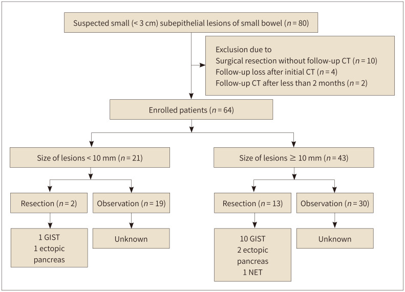
Materials and methods: Consecutive patients who were suspected of having subepithelial tumors of the small bowel between January 2005 and December 2020 were reviewed. Eligible patients with suspected small (< 30 mm) subepithelial tumors on at least two CT evaluations were included in the analysis. The patients' data on demographic characteristics, tumoral characteristics, and tumoral size changes during the follow-up were collected.
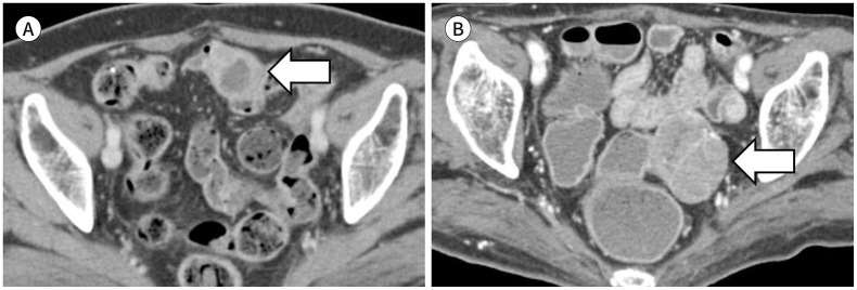
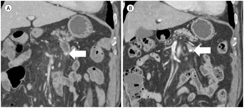
Results: This study included 64 patients with suspected small subepithelial tumors (n = 64) of the small bowel. After a median follow-up of 15.8 months, the diameter and volume growth rates were 0.02 mm/month and 1.5 mm3/month, respectively. A significant correlation was observed between the initial size and the growth rate of the small bowel subepithelial tumors. The group of large-sized tumors (initial diameter ≥ 10 mm) tended to show lobulated contours, heterogeneous enhancement, and necrotic changes more frequently than the group of small-sized tumors (initial diameter < 10 mm).
Conclusion: Small bowel subepithelial tumors measuring less than 10 mm grew more slowly than those measuring 10-30 mm.

 求助内容:
求助内容: 应助结果提醒方式:


