So Jung Ki, Chul Hwan Park, Kyunghwa Han, Jae Min Shin, Ji Young Kim, Tae Hoon Kim
{"title":"Utility of the 16-cm Axial Volume Scan Technique for Coronary Artery Calcium Scoring on Non-Enhanced Chest CT: A Prospective Pilot Study.","authors":"So Jung Ki, Chul Hwan Park, Kyunghwa Han, Jae Min Shin, Ji Young Kim, Tae Hoon Kim","doi":"10.3348/jksr.2020.0156","DOIUrl":null,"url":null,"abstract":"<p><strong>Purpose: </strong>This study aimed to evaluate the utility of the 16-cm axial volume scan technique for calculating the coronary artery calcium score (CACS) using non-enhanced chest CT.</p><p><strong>Materials and methods: </strong>This study prospectively enrolled 20 participants who underwent both, non-enhanced chest CT (16-cm-coverage axial volume scan technique) and calcium-score CT, with the same parameters, differing only in slice thickness (in non-enhanced chest CT = 0.625, 1.25, 2.5 mm; in calcium score CT = 2.5 mm). The CACS was calculated using the conventional Agatston method. The difference between the CACS obtained from the two CT scans was compared, and the degree of agreement for the clinical significance of the CACS was confirmed through sectional analysis. Each calcified lesion was classified by location and size, and a one-to-one comparison of non-contrast-enhanced chest CT and calcium score CT was performed.</p><p><strong>Results: </strong>The correlation coefficients of the CACS obtained from the two CT scans for slice thickness of 2.5, 1.25, and 0.625 mm were 0.9850, 0.9688, and 0.9834, respectively. The mean differences between the CACS were -21.4% at 0.625 mm, -39.4% at 1.25 mm, and -76.2% at 2.5 mm slice thicknesses. Sectional analysis revealed that 16 (80%), 16 (80%), and 13 (65%) patients showed agreement for the degree of coronary artery disease at each slice interval, respectively. Inter-reader agreement was high for each slice interval. The 0.625 mm CT showed the highest sensitivity for detecting calcified lesions.</p><p><strong>Conclusion: </strong>The values in the non-contrast-enhanced chest CT, using the 16-cm axial volume scan technique, were similar to those obtained using the CACS in the calcium score CT, at 0.625 mm slice thickness without electrocardiogram gating. This can ultimately help predict cardiovascular risk without additional radiation exposure.</p>","PeriodicalId":74904,"journal":{"name":"Taehan Yongsang Uihakhoe chi","volume":"82 6","pages":"1493-1504"},"PeriodicalIF":0.0000,"publicationDate":"2021-11-01","publicationTypes":"Journal Article","fieldsOfStudy":null,"isOpenAccess":false,"openAccessPdf":"https://ftp.ncbi.nlm.nih.gov/pub/pmc/oa_pdf/be/bf/jksr-82-1493.PMC9431984.pdf","citationCount":"0","resultStr":null,"platform":"Semanticscholar","paperid":null,"PeriodicalName":"Taehan Yongsang Uihakhoe chi","FirstCategoryId":"1085","ListUrlMain":"https://doi.org/10.3348/jksr.2020.0156","RegionNum":0,"RegionCategory":null,"ArticlePicture":[],"TitleCN":null,"AbstractTextCN":null,"PMCID":null,"EPubDate":"2021/8/27 0:00:00","PubModel":"Epub","JCR":"","JCRName":"","Score":null,"Total":0}
引用次数: 0
Abstract
Purpose: This study aimed to evaluate the utility of the 16-cm axial volume scan technique for calculating the coronary artery calcium score (CACS) using non-enhanced chest CT.
Materials and methods: This study prospectively enrolled 20 participants who underwent both, non-enhanced chest CT (16-cm-coverage axial volume scan technique) and calcium-score CT, with the same parameters, differing only in slice thickness (in non-enhanced chest CT = 0.625, 1.25, 2.5 mm; in calcium score CT = 2.5 mm). The CACS was calculated using the conventional Agatston method. The difference between the CACS obtained from the two CT scans was compared, and the degree of agreement for the clinical significance of the CACS was confirmed through sectional analysis. Each calcified lesion was classified by location and size, and a one-to-one comparison of non-contrast-enhanced chest CT and calcium score CT was performed.
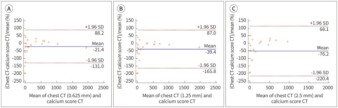
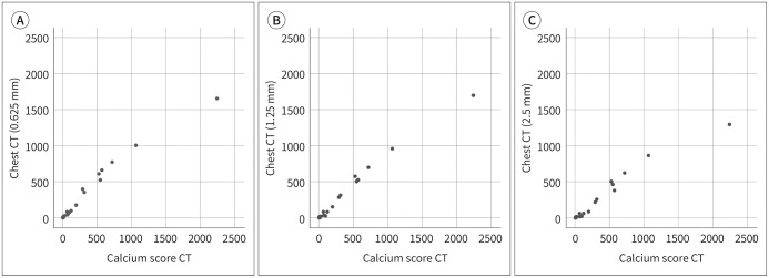
Results: The correlation coefficients of the CACS obtained from the two CT scans for slice thickness of 2.5, 1.25, and 0.625 mm were 0.9850, 0.9688, and 0.9834, respectively. The mean differences between the CACS were -21.4% at 0.625 mm, -39.4% at 1.25 mm, and -76.2% at 2.5 mm slice thicknesses. Sectional analysis revealed that 16 (80%), 16 (80%), and 13 (65%) patients showed agreement for the degree of coronary artery disease at each slice interval, respectively. Inter-reader agreement was high for each slice interval. The 0.625 mm CT showed the highest sensitivity for detecting calcified lesions.
Conclusion: The values in the non-contrast-enhanced chest CT, using the 16-cm axial volume scan technique, were similar to those obtained using the CACS in the calcium score CT, at 0.625 mm slice thickness without electrocardiogram gating. This can ultimately help predict cardiovascular risk without additional radiation exposure.

 求助内容:
求助内容: 应助结果提醒方式:


