Ji Yeon Park, Seong Yoon Yi, Ji Young Lee, Tae Jung Kwon
{"title":"A Case Report of Axillary Hibernoma: US, CT, MR, and Histopathologic Findings.","authors":"Ji Yeon Park, Seong Yoon Yi, Ji Young Lee, Tae Jung Kwon","doi":"10.3348/jksr.2021.0030","DOIUrl":null,"url":null,"abstract":"<p><p>Hibernoma is a rare benign tumor of brown adipose tissue. Herein, we report a case of axillary hibernoma in a 53-year-old female and discuss the various radiologic findings. The US revealed a 4.5-cm well-defined oval heterogenous hyperechoic mass in the right axilla with anterior displacement of the axillary vessels. Non-enhanced chest CT showed a 5.0-cm well defined, oval, and low-attenuated mass. MRI demonstrated a 5.5-cm mass with heterogeneous intermediate-to-high signal intensity on T1-and T2-weighted images and irregular enhancement at the peripheral portion. The patient underwent an US-guided core needle biopsy and the final diagnosis was hibernoma.</p>","PeriodicalId":74904,"journal":{"name":"Taehan Yongsang Uihakhoe chi","volume":" ","pages":"439-443"},"PeriodicalIF":0.0000,"publicationDate":"2022-03-01","publicationTypes":"Journal Article","fieldsOfStudy":null,"isOpenAccess":false,"openAccessPdf":"https://ftp.ncbi.nlm.nih.gov/pub/pmc/oa_pdf/f3/28/jksr-83-439.PMC9514428.pdf","citationCount":"0","resultStr":null,"platform":"Semanticscholar","paperid":null,"PeriodicalName":"Taehan Yongsang Uihakhoe chi","FirstCategoryId":"1085","ListUrlMain":"https://doi.org/10.3348/jksr.2021.0030","RegionNum":0,"RegionCategory":null,"ArticlePicture":[],"TitleCN":null,"AbstractTextCN":null,"PMCID":null,"EPubDate":"2021/10/18 0:00:00","PubModel":"Epub","JCR":"","JCRName":"","Score":null,"Total":0}
引用次数: 0
Abstract
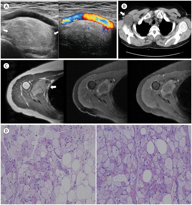
Hibernoma is a rare benign tumor of brown adipose tissue. Herein, we report a case of axillary hibernoma in a 53-year-old female and discuss the various radiologic findings. The US revealed a 4.5-cm well-defined oval heterogenous hyperechoic mass in the right axilla with anterior displacement of the axillary vessels. Non-enhanced chest CT showed a 5.0-cm well defined, oval, and low-attenuated mass. MRI demonstrated a 5.5-cm mass with heterogeneous intermediate-to-high signal intensity on T1-and T2-weighted images and irregular enhancement at the peripheral portion. The patient underwent an US-guided core needle biopsy and the final diagnosis was hibernoma.

 求助内容:
求助内容: 应助结果提醒方式:


