Sung Hyun Yu, Young Sup Shim, So Hyun Park, Seung Joon Choi, Dong Hae Chung, Sang Jin Yoon
{"title":"MRI Findings of Renal Myxoma: A Case Report and Literature Review.","authors":"Sung Hyun Yu, Young Sup Shim, So Hyun Park, Seung Joon Choi, Dong Hae Chung, Sang Jin Yoon","doi":"10.3348/jksr.2019.0174","DOIUrl":null,"url":null,"abstract":"<p><p>Renal myxomas are very rare benign tumors. To date, a few cases have been reported in English literature, mostly in pathology and urology journals. Thus, there are few reports on the radiological findings associated with renal myxomas. We report on the imaging findings in a case of renal myxoma in a 62-year-old male. MRI demonstrated a well-defined mass in the left renal sinus, with intermediate high signal intensity on T2-weighted images and low signal intensity on T1-weighted images. The tumor showed gradual enhancement on contrast-enhanced T1-weighted images.</p>","PeriodicalId":74904,"journal":{"name":"Taehan Yongsang Uihakhoe chi","volume":" ","pages":"162-167"},"PeriodicalIF":0.0000,"publicationDate":"2022-01-01","publicationTypes":"Journal Article","fieldsOfStudy":null,"isOpenAccess":false,"openAccessPdf":"https://ftp.ncbi.nlm.nih.gov/pub/pmc/oa_pdf/89/f4/jksr-83-162.PMC9238218.pdf","citationCount":"0","resultStr":null,"platform":"Semanticscholar","paperid":null,"PeriodicalName":"Taehan Yongsang Uihakhoe chi","FirstCategoryId":"1085","ListUrlMain":"https://doi.org/10.3348/jksr.2019.0174","RegionNum":0,"RegionCategory":null,"ArticlePicture":[],"TitleCN":null,"AbstractTextCN":null,"PMCID":null,"EPubDate":"2022/1/21 0:00:00","PubModel":"Epub","JCR":"","JCRName":"","Score":null,"Total":0}
引用次数: 0
Abstract
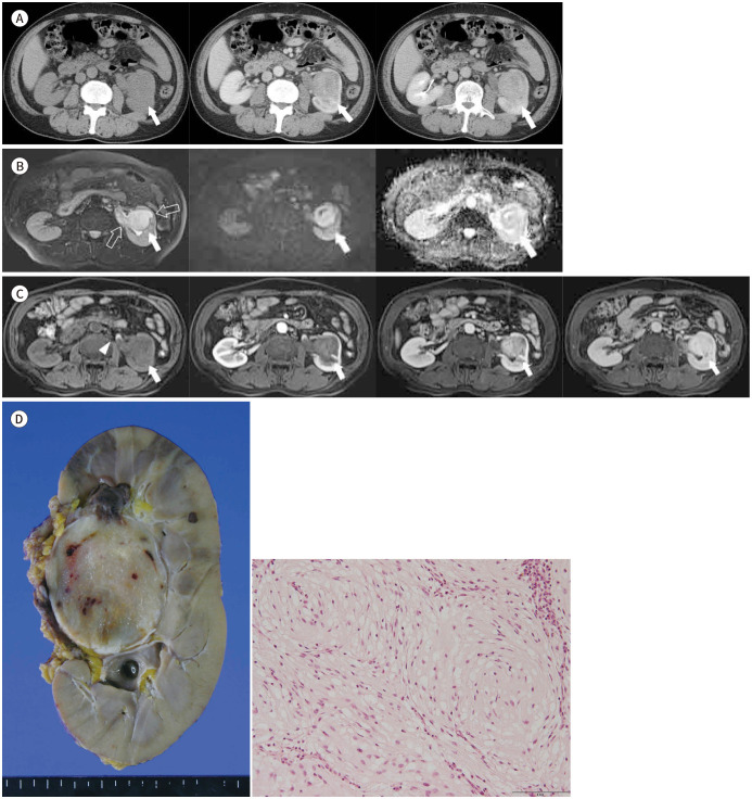
Renal myxomas are very rare benign tumors. To date, a few cases have been reported in English literature, mostly in pathology and urology journals. Thus, there are few reports on the radiological findings associated with renal myxomas. We report on the imaging findings in a case of renal myxoma in a 62-year-old male. MRI demonstrated a well-defined mass in the left renal sinus, with intermediate high signal intensity on T2-weighted images and low signal intensity on T1-weighted images. The tumor showed gradual enhancement on contrast-enhanced T1-weighted images.

 求助内容:
求助内容: 应助结果提醒方式:


