Nabil K El Ayoubi, Hadi M Sabbagh, Nicole Bou Rjeily, Salem Hannoun, Samia J Khoury
{"title":"Rate of Retinal Layer Thinning as a Biomarker for Conversion to Progressive Disease in Multiple Sclerosis.","authors":"Nabil K El Ayoubi, Hadi M Sabbagh, Nicole Bou Rjeily, Salem Hannoun, Samia J Khoury","doi":"10.1212/NXI.0000000000200030","DOIUrl":null,"url":null,"abstract":"<p><strong>Background and objectives: </strong>The diagnosis of secondary progressive multiple sclerosis (SPMS) is often delayed because of the lack of objective clinical tools, which increases the diagnostic uncertainty and hampers the therapeutic development in progressive multiple sclerosis (MS). Optical coherence tomography (OCT) has been proposed as a promising biomarker of progressive neurodegeneration. To explore longitudinal changes in the thicknesses of retinal layers on OCT in individuals with relapsing-remitting MS (RRMS) who converted to SPMS vs matched patients with RRMS who did not convert to SPMS. Our hypothesis is that the 2 cohorts exhibit different rates of retinal thinning.</p><p><strong>Methods: </strong>From our prospective observational cohort of patients with MS at the American University of Beirut, we selected patients with RRMS who converted to SPMS during the observation period and patients with RRMS, matched by age, disease duration, and Expanded Disability Status Scale (EDSS) at the first visit. Baseline retinal measurements were obtained using spectral domain OCT, and all patients underwent clinical and OCT evaluation every 6-12 months on average throughout the study period (mean = 4 years). Mixed-effect regression models were used to assess the annualized rates of retinal changes and the differences between the 2 groups and between converters to SPMS before and after their conversion.</p><p><strong>Results: </strong>A total of 61 participants were selected (21 SPMS and 40 RRMS). There were no differences in baseline characteristics and retinal measurements between the 2 groups. The annualized rates of thinning of all retinal layers, except for macular volume, were greater in converters before conversion compared with nonconverters by 112% for peripapillary retinal nerve fiber layer (<i>p</i> = 0.008), 344% for tRNFL (<i>p</i> < 0.0001), and 82% for cell-inner plexiform layer (GCIPL) (<i>p</i> = 0.002). When comparing the annualized rate of thinning for the same patients with SPMS before and after conversion, no significant differences were found except for tRNFL and GCIPL with slower thinning rates postconversion (46% and 68%, respectively).</p><p><strong>Discussion: </strong>Patients who converted to SPMS exhibited faster retinal thinning as reflected on OCT. Longitudinal assessment of retinal thinning could confirm the transition to SPMS and help with the therapeutic decision making for patients with MS with clinical suspicion of disease progression.</p>","PeriodicalId":520720,"journal":{"name":"Neurology(R) neuroimmunology & neuroinflammation","volume":" ","pages":""},"PeriodicalIF":7.5000,"publicationDate":"2022-10-13","publicationTypes":"Journal Article","fieldsOfStudy":null,"isOpenAccess":false,"openAccessPdf":"https://ftp.ncbi.nlm.nih.gov/pub/pmc/oa_pdf/e4/24/NXI-2022-200036.PMC9562042.pdf","citationCount":"0","resultStr":null,"platform":"Semanticscholar","paperid":null,"PeriodicalName":"Neurology(R) neuroimmunology & neuroinflammation","FirstCategoryId":"3","ListUrlMain":"https://doi.org/10.1212/NXI.0000000000200030","RegionNum":0,"RegionCategory":null,"ArticlePicture":[],"TitleCN":null,"AbstractTextCN":null,"PMCID":null,"EPubDate":"2022/11/1 0:00:00","PubModel":"Print","JCR":"","JCRName":"","Score":null,"Total":0}
引用次数: 0
Abstract
Background and objectives: The diagnosis of secondary progressive multiple sclerosis (SPMS) is often delayed because of the lack of objective clinical tools, which increases the diagnostic uncertainty and hampers the therapeutic development in progressive multiple sclerosis (MS). Optical coherence tomography (OCT) has been proposed as a promising biomarker of progressive neurodegeneration. To explore longitudinal changes in the thicknesses of retinal layers on OCT in individuals with relapsing-remitting MS (RRMS) who converted to SPMS vs matched patients with RRMS who did not convert to SPMS. Our hypothesis is that the 2 cohorts exhibit different rates of retinal thinning.
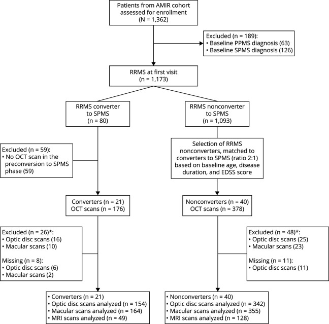
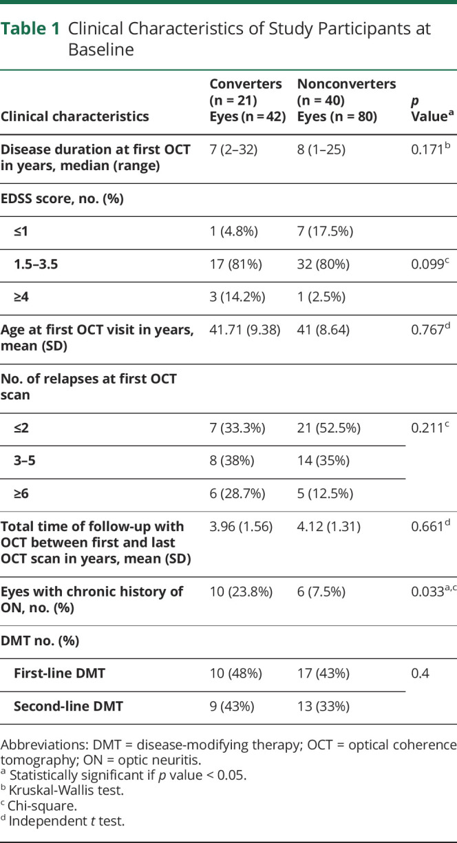
Methods: From our prospective observational cohort of patients with MS at the American University of Beirut, we selected patients with RRMS who converted to SPMS during the observation period and patients with RRMS, matched by age, disease duration, and Expanded Disability Status Scale (EDSS) at the first visit. Baseline retinal measurements were obtained using spectral domain OCT, and all patients underwent clinical and OCT evaluation every 6-12 months on average throughout the study period (mean = 4 years). Mixed-effect regression models were used to assess the annualized rates of retinal changes and the differences between the 2 groups and between converters to SPMS before and after their conversion.
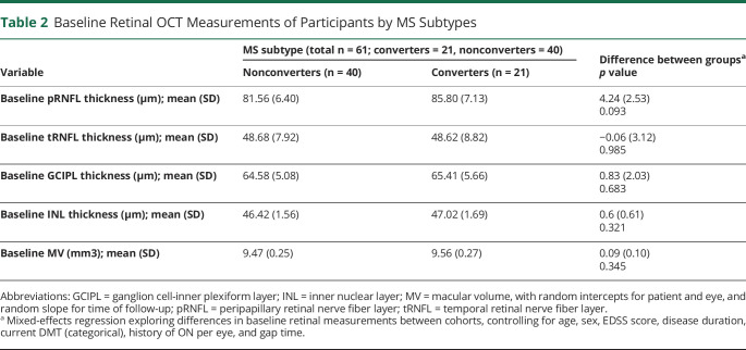
Results: A total of 61 participants were selected (21 SPMS and 40 RRMS). There were no differences in baseline characteristics and retinal measurements between the 2 groups. The annualized rates of thinning of all retinal layers, except for macular volume, were greater in converters before conversion compared with nonconverters by 112% for peripapillary retinal nerve fiber layer (p = 0.008), 344% for tRNFL (p < 0.0001), and 82% for cell-inner plexiform layer (GCIPL) (p = 0.002). When comparing the annualized rate of thinning for the same patients with SPMS before and after conversion, no significant differences were found except for tRNFL and GCIPL with slower thinning rates postconversion (46% and 68%, respectively).
Discussion: Patients who converted to SPMS exhibited faster retinal thinning as reflected on OCT. Longitudinal assessment of retinal thinning could confirm the transition to SPMS and help with the therapeutic decision making for patients with MS with clinical suspicion of disease progression.

 求助内容:
求助内容: 应助结果提醒方式:


