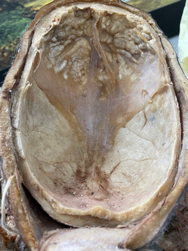
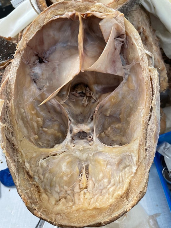
{"title":"Hyperostosis Frontalis Interna and a Question on Its Pathology: A Case Report.","authors":"Jennifer Michelle Stiene, Patrick William Frank","doi":"10.12659/AJCR.937450","DOIUrl":null,"url":null,"abstract":"<p><p>BACKGROUND Hyperostosis frontalis interna is a boney overgrowth of the inner side of the frontal bone of the skull caused by overgrowth of the endocranial surface. It is most often found in women after menopause. It is also associated with hormonal imbalance, being overweight, history of headaches, and neurocognitive degenerative conditions. Female gender, advanced age, extended estrogen stimulation, and elevated leptin levels may also play a role. The thickening is usually confined to the frontal bone, but it can spread as far as the anterior parietal and temporal bones. CASE REPORT During a medical school dissection course, an extensive boney overgrowth in the frontal regions covering the inside of the frontal bone of the skull of a 90-year-old female donor, who died of a cerebrovascular infarction, was identified. This boney overgrowth was mainly confined within the frontal region, but there was some boney overgrowth that extended to the temporal bones. The overgrowth in the endocranium of the temporal bone was not as severe as the overgrowth of the frontal bone. The morphology of the overgrowth was rigid, uneven, and bumpy. Based upon the physical characteristics, we concluded that this presentation was consistent with hyperostosis frontalis interna. CONCLUSIONS Our female donor was found to exhibit a phenomenon which could be clinically underdiagnosed due to its internal nature and asymptomatic presentation. Insight into the potential causes of HFI and its identification during clinical evaluation offers a path for future research to better identify and manage cases of HFI.</p>","PeriodicalId":205256,"journal":{"name":"The American Journal of Case Reports","volume":" ","pages":"e937450"},"PeriodicalIF":0.0000,"publicationDate":"2022-10-11","publicationTypes":"Journal Article","fieldsOfStudy":null,"isOpenAccess":false,"openAccessPdf":"https://ftp.ncbi.nlm.nih.gov/pub/pmc/oa_pdf/98/5b/amjcaserep-23-e937450.PMC9575136.pdf","citationCount":"1","resultStr":null,"platform":"Semanticscholar","paperid":null,"PeriodicalName":"The American Journal of Case Reports","FirstCategoryId":"1085","ListUrlMain":"https://doi.org/10.12659/AJCR.937450","RegionNum":0,"RegionCategory":null,"ArticlePicture":[],"TitleCN":null,"AbstractTextCN":null,"PMCID":null,"EPubDate":"","PubModel":"","JCR":"","JCRName":"","Score":null,"Total":0}
引用次数: 1
Abstract
BACKGROUND Hyperostosis frontalis interna is a boney overgrowth of the inner side of the frontal bone of the skull caused by overgrowth of the endocranial surface. It is most often found in women after menopause. It is also associated with hormonal imbalance, being overweight, history of headaches, and neurocognitive degenerative conditions. Female gender, advanced age, extended estrogen stimulation, and elevated leptin levels may also play a role. The thickening is usually confined to the frontal bone, but it can spread as far as the anterior parietal and temporal bones. CASE REPORT During a medical school dissection course, an extensive boney overgrowth in the frontal regions covering the inside of the frontal bone of the skull of a 90-year-old female donor, who died of a cerebrovascular infarction, was identified. This boney overgrowth was mainly confined within the frontal region, but there was some boney overgrowth that extended to the temporal bones. The overgrowth in the endocranium of the temporal bone was not as severe as the overgrowth of the frontal bone. The morphology of the overgrowth was rigid, uneven, and bumpy. Based upon the physical characteristics, we concluded that this presentation was consistent with hyperostosis frontalis interna. CONCLUSIONS Our female donor was found to exhibit a phenomenon which could be clinically underdiagnosed due to its internal nature and asymptomatic presentation. Insight into the potential causes of HFI and its identification during clinical evaluation offers a path for future research to better identify and manage cases of HFI.

 求助内容:
求助内容: 应助结果提醒方式:


