Lara Nikel, Magdalena W Sliwinska, Emel Kucuk, Leslie G Ungerleider, David Pitcher
{"title":"Measuring the response to visually presented faces in the human lateral prefrontal cortex.","authors":"Lara Nikel, Magdalena W Sliwinska, Emel Kucuk, Leslie G Ungerleider, David Pitcher","doi":"10.1093/texcom/tgac036","DOIUrl":null,"url":null,"abstract":"<p><p>Neuroimaging studies identify multiple face-selective areas in the human brain. In the current study, we compared the functional response of the face area in the lateral prefrontal cortex to that of other face-selective areas. In Experiment 1, participants (<i>n</i> = 32) were scanned viewing videos containing faces, bodies, scenes, objects, and scrambled objects. We identified a face-selective area in the right inferior frontal gyrus (rIFG). In Experiment 2, participants (<i>n</i> = 24) viewed the same videos or static images. Results showed that the rIFG, right posterior superior temporal sulcus (rpSTS), and right occipital face area (rOFA) exhibited a greater response to moving than static faces. In Experiment 3, participants (<i>n</i> = 18) viewed face videos in the contralateral and ipsilateral visual fields. Results showed that the rIFG and rpSTS showed no visual field bias, while the rOFA and right fusiform face area (rFFA) showed a contralateral bias. These experiments suggest two conclusions; firstly, in all three experiments, the face area in the IFG was not as reliably identified as face areas in the occipitotemporal cortex. Secondly, the similarity of the response profiles in the IFG and pSTS suggests the areas may perform similar cognitive functions, a conclusion consistent with prior neuroanatomical and functional connectivity evidence.</p>","PeriodicalId":72551,"journal":{"name":"Cerebral cortex communications","volume":" ","pages":"tgac036"},"PeriodicalIF":0.0000,"publicationDate":"2022-08-18","publicationTypes":"Journal Article","fieldsOfStudy":null,"isOpenAccess":false,"openAccessPdf":"https://www.ncbi.nlm.nih.gov/pmc/articles/PMC9491845/pdf/","citationCount":"6","resultStr":null,"platform":"Semanticscholar","paperid":null,"PeriodicalName":"Cerebral cortex communications","FirstCategoryId":"1085","ListUrlMain":"https://doi.org/10.1093/texcom/tgac036","RegionNum":0,"RegionCategory":null,"ArticlePicture":[],"TitleCN":null,"AbstractTextCN":null,"PMCID":null,"EPubDate":"2022/1/1 0:00:00","PubModel":"eCollection","JCR":"","JCRName":"","Score":null,"Total":0}
引用次数: 6
Abstract
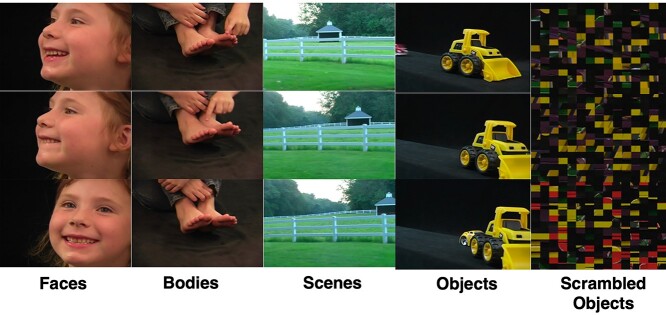
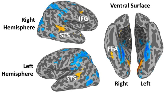
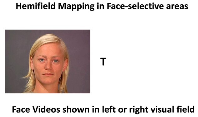
Neuroimaging studies identify multiple face-selective areas in the human brain. In the current study, we compared the functional response of the face area in the lateral prefrontal cortex to that of other face-selective areas. In Experiment 1, participants (n = 32) were scanned viewing videos containing faces, bodies, scenes, objects, and scrambled objects. We identified a face-selective area in the right inferior frontal gyrus (rIFG). In Experiment 2, participants (n = 24) viewed the same videos or static images. Results showed that the rIFG, right posterior superior temporal sulcus (rpSTS), and right occipital face area (rOFA) exhibited a greater response to moving than static faces. In Experiment 3, participants (n = 18) viewed face videos in the contralateral and ipsilateral visual fields. Results showed that the rIFG and rpSTS showed no visual field bias, while the rOFA and right fusiform face area (rFFA) showed a contralateral bias. These experiments suggest two conclusions; firstly, in all three experiments, the face area in the IFG was not as reliably identified as face areas in the occipitotemporal cortex. Secondly, the similarity of the response profiles in the IFG and pSTS suggests the areas may perform similar cognitive functions, a conclusion consistent with prior neuroanatomical and functional connectivity evidence.

 求助内容:
求助内容: 应助结果提醒方式:


