Lucas Alves Teixeira Oliveira, Rhayane Patrícia Rodrigues Oliveira, Laura Cardoso Vasconcelos, Achilles Alves de Levy Machado, Gustavo Lara Rezende, Fayez Bahmad
{"title":"A Rare Case of Giant Cavernous Hemangioma of the Maxillary Sinus.","authors":"Lucas Alves Teixeira Oliveira, Rhayane Patrícia Rodrigues Oliveira, Laura Cardoso Vasconcelos, Achilles Alves de Levy Machado, Gustavo Lara Rezende, Fayez Bahmad","doi":"10.12659/AJCR.937191","DOIUrl":null,"url":null,"abstract":"<p><p>BACKGROUND Hemangiomas are commonly located in the head and neck and rarely in the paranasal sinuses. These are benign vascular lesions, but with an increased risk of bleeding. The surgical approach must have detailed prior planning, given the increased risk of intraoperative bleeding. We herein describe the case of a 32-year-old male patient with recurrent epistaxis, nasal obstruction, and facial deformity due to a giant cavernous hemangioma successfully treated by endoscopic sinus surgery. CASE REPORT A 32-year-old man had nasal obstruction and intermittent epistaxis for 2 months. Physical examination also revealed facial deformity with enlargement of the nasal base and bulging in the maxillary region on the right. A soft and friable lesion occupying the entire right nasal cavity without bone erosion was observed on computed tomography (CT scan). Before surgery, the patient underwent angiographic evaluation, with evidence of main irrigation of the lesion by the right maxillary artery, which was then embolized. The patient underwent endoscopic nasal surgery. He maintained postoperative follow-up for 18 months, without recurrence of the lesion. Anatomopathological examination confirmed a cavernous hemangioma. CONCLUSIONS Cavernous hemangioma is a benign lesion of the paranasal sinuses. Due to non-specific clinical and radiological findings, its preoperative diagnosis is always challenging. The high index of suspicion of the malignancy should only be discarded after complete anatomopathological evaluation. A correct diagnosis is essential to avoid facial anatomical remodeling while excluding the diagnosis of other malignant lesions.</p>","PeriodicalId":205256,"journal":{"name":"The American Journal of Case Reports","volume":" ","pages":"e937191"},"PeriodicalIF":0.0000,"publicationDate":"2022-10-09","publicationTypes":"Journal Article","fieldsOfStudy":null,"isOpenAccess":false,"openAccessPdf":"https://ftp.ncbi.nlm.nih.gov/pub/pmc/oa_pdf/8e/69/amjcaserep-23-e937191.PMC9557247.pdf","citationCount":"0","resultStr":null,"platform":"Semanticscholar","paperid":null,"PeriodicalName":"The American Journal of Case Reports","FirstCategoryId":"1085","ListUrlMain":"https://doi.org/10.12659/AJCR.937191","RegionNum":0,"RegionCategory":null,"ArticlePicture":[],"TitleCN":null,"AbstractTextCN":null,"PMCID":null,"EPubDate":"","PubModel":"","JCR":"","JCRName":"","Score":null,"Total":0}
引用次数: 0
Abstract
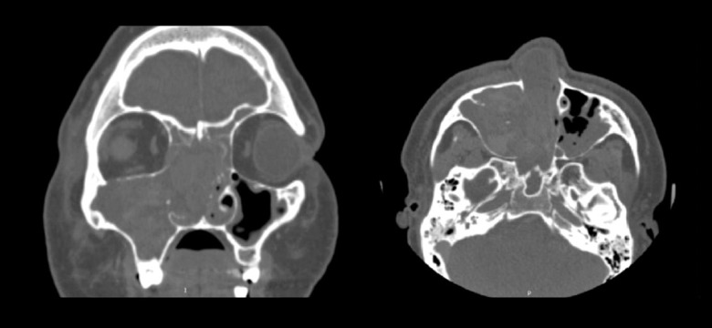
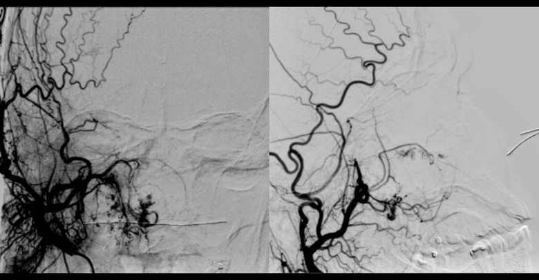
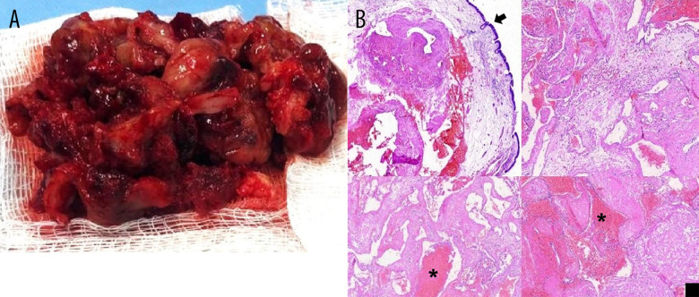
BACKGROUND Hemangiomas are commonly located in the head and neck and rarely in the paranasal sinuses. These are benign vascular lesions, but with an increased risk of bleeding. The surgical approach must have detailed prior planning, given the increased risk of intraoperative bleeding. We herein describe the case of a 32-year-old male patient with recurrent epistaxis, nasal obstruction, and facial deformity due to a giant cavernous hemangioma successfully treated by endoscopic sinus surgery. CASE REPORT A 32-year-old man had nasal obstruction and intermittent epistaxis for 2 months. Physical examination also revealed facial deformity with enlargement of the nasal base and bulging in the maxillary region on the right. A soft and friable lesion occupying the entire right nasal cavity without bone erosion was observed on computed tomography (CT scan). Before surgery, the patient underwent angiographic evaluation, with evidence of main irrigation of the lesion by the right maxillary artery, which was then embolized. The patient underwent endoscopic nasal surgery. He maintained postoperative follow-up for 18 months, without recurrence of the lesion. Anatomopathological examination confirmed a cavernous hemangioma. CONCLUSIONS Cavernous hemangioma is a benign lesion of the paranasal sinuses. Due to non-specific clinical and radiological findings, its preoperative diagnosis is always challenging. The high index of suspicion of the malignancy should only be discarded after complete anatomopathological evaluation. A correct diagnosis is essential to avoid facial anatomical remodeling while excluding the diagnosis of other malignant lesions.

 求助内容:
求助内容: 应助结果提醒方式:


