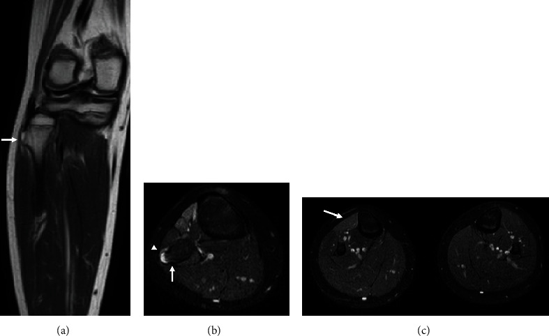
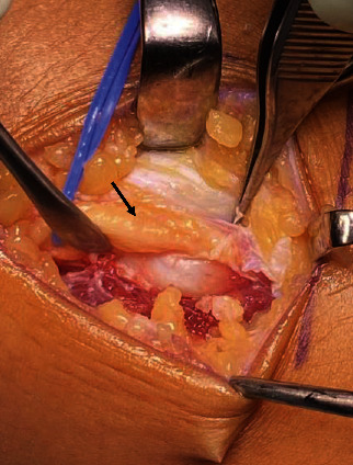
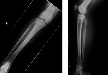
{"title":"Peroneal Nerve Palsy Caused by Proximal Fibular Solitary Osteochondroma: Case Report and Literature Review.","authors":"Takashi Kozu, Masayoshi Machida, Katsuaki Taira, Noboru Oikawa, Naho Nemoto, Kazuyoshi Nakanishi","doi":"10.1155/2022/5865040","DOIUrl":null,"url":null,"abstract":"<p><p>Osteochondroma is a relatively common benign tumor of the bone, and compressive neuropathies due to osteochondroma are comparatively rare. Here, we present a rare case of osteochondroma of the fibular head that caused peroneal nerve palsy in an 8-year-old girl. Physical examination revealed 0/5 tibialis anterior, 1/5 extensor hallucis longus, and 1/5 peroneal brevis muscle power-according to the manual muscle testing grading system, as well as numbness on the lateral side of the right leg and the back of the foot. Radiological examination and ultrasound revealed a bone tumor in the head of the right fibula. Magnetic resonance imaging ruled out spinal nerve root compression. It was discovered that the bone tumor in the fibular head had compressed and displaced the common peroneal nerve. The patient underwent surgical decompression of the right peroneal nerve. A bone region measuring 22 × 14 × 8 mm was removed. Three months postoperatively, the preoperative neurological deficits were found to be nearly resolved. The patient presented with a foot drop for 1 year, but symptoms resolved 3 months after surgery. Conventional wisdom states that surgery should be performed within 3 months, but we recommend that surgery be performed as soon as diagnosis is made even in cases with a long history, as it may improve patient symptoms and outcomes.</p>","PeriodicalId":30287,"journal":{"name":"Case Reports in Orthopedics","volume":" ","pages":"5865040"},"PeriodicalIF":0.6000,"publicationDate":"2022-09-13","publicationTypes":"Journal Article","fieldsOfStudy":null,"isOpenAccess":false,"openAccessPdf":"https://www.ncbi.nlm.nih.gov/pmc/articles/PMC9489407/pdf/","citationCount":"1","resultStr":null,"platform":"Semanticscholar","paperid":null,"PeriodicalName":"Case Reports in Orthopedics","FirstCategoryId":"1085","ListUrlMain":"https://doi.org/10.1155/2022/5865040","RegionNum":0,"RegionCategory":null,"ArticlePicture":[],"TitleCN":null,"AbstractTextCN":null,"PMCID":null,"EPubDate":"2022/1/1 0:00:00","PubModel":"eCollection","JCR":"Q4","JCRName":"ORTHOPEDICS","Score":null,"Total":0}
引用次数: 1
Abstract
Osteochondroma is a relatively common benign tumor of the bone, and compressive neuropathies due to osteochondroma are comparatively rare. Here, we present a rare case of osteochondroma of the fibular head that caused peroneal nerve palsy in an 8-year-old girl. Physical examination revealed 0/5 tibialis anterior, 1/5 extensor hallucis longus, and 1/5 peroneal brevis muscle power-according to the manual muscle testing grading system, as well as numbness on the lateral side of the right leg and the back of the foot. Radiological examination and ultrasound revealed a bone tumor in the head of the right fibula. Magnetic resonance imaging ruled out spinal nerve root compression. It was discovered that the bone tumor in the fibular head had compressed and displaced the common peroneal nerve. The patient underwent surgical decompression of the right peroneal nerve. A bone region measuring 22 × 14 × 8 mm was removed. Three months postoperatively, the preoperative neurological deficits were found to be nearly resolved. The patient presented with a foot drop for 1 year, but symptoms resolved 3 months after surgery. Conventional wisdom states that surgery should be performed within 3 months, but we recommend that surgery be performed as soon as diagnosis is made even in cases with a long history, as it may improve patient symptoms and outcomes.

 求助内容:
求助内容: 应助结果提醒方式:


