Transtibial Pullout Repair of Lateral Meniscus Posterior Root Tear with Tissue Loss: A Case with Anterior Cruciate Ligament Injury and Medial Meniscus Tear.
{"title":"Transtibial Pullout Repair of Lateral Meniscus Posterior Root Tear with Tissue Loss: A Case with Anterior Cruciate Ligament Injury and Medial Meniscus Tear.","authors":"Masanori Tamura, Takayuki Furumatsu, Takaaki Hiranaka, Keisuke Kintaka, Naohiro Higashihara, Yusuke Kamatsuki, Eiji Nakata, Toshifumi Ozaki","doi":"10.1155/2022/9776388","DOIUrl":null,"url":null,"abstract":"<p><p>Lateral meniscus (LM) posterior root tear (LMPRT) is mainly caused by trauma, especially trauma associated with anterior cruciate ligament (ACL) injuries. Although a transtibial pullout repair or a side-to-side repair is commonly performed for LMPRT, to the best of our knowledge, there is no clinical report of LMPRT with tissue loss using the pullout technique. Thus, the purpose of this report was to describe a clinical, radiographic, and arthroscopic outcome after pullout repair for a case of LMPRT with a large defect with a chronic ACL tear and complex medial meniscus (MM) tears. A 31-year-old man complained of knee pain and restricted range of motion after twisting his knee when he stepped on an iron pipe. The patient had a football-related injury to his right knee 14 years before presentation, and since then, the patient's knee has given out more than 10 times but was left unassessed. Magnetic resonance imaging showed LMPRT with tissue loss, ACL tears, and complex MM tears. Transtibial pullout repair of the LMPRT with ACL reconstruction and MM repairs were performed. Following the pullout repair of the LMPRT, an approximately 6 mm gap remained between the LM posterior root and root insertion. However, magnetic resonance imaging and second-look arthroscopy at 1 year postoperatively revealed meniscal healing, gap filling with some regeneration tissue, of the LM posterior root. Furthermore, the lateral meniscus extrusion in the coronal plane improved from 3.1 mm (preoperative) to 1.6 mm (1 year postoperatively). Transtibial pullout repair with the remaining gap could be a viable treatment option for LMPRT with tissue loss, combined with ACL reconstruction.</p>","PeriodicalId":30287,"journal":{"name":"Case Reports in Orthopedics","volume":" ","pages":"9776388"},"PeriodicalIF":0.6000,"publicationDate":"2022-08-31","publicationTypes":"Journal Article","fieldsOfStudy":null,"isOpenAccess":false,"openAccessPdf":"https://www.ncbi.nlm.nih.gov/pmc/articles/PMC9453023/pdf/","citationCount":"0","resultStr":null,"platform":"Semanticscholar","paperid":null,"PeriodicalName":"Case Reports in Orthopedics","FirstCategoryId":"1085","ListUrlMain":"https://doi.org/10.1155/2022/9776388","RegionNum":0,"RegionCategory":null,"ArticlePicture":[],"TitleCN":null,"AbstractTextCN":null,"PMCID":null,"EPubDate":"2022/1/1 0:00:00","PubModel":"eCollection","JCR":"Q4","JCRName":"ORTHOPEDICS","Score":null,"Total":0}
引用次数: 0
Abstract
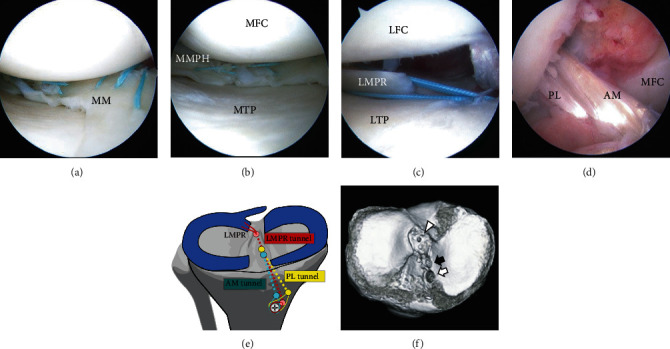
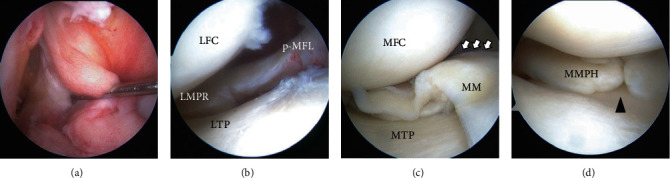
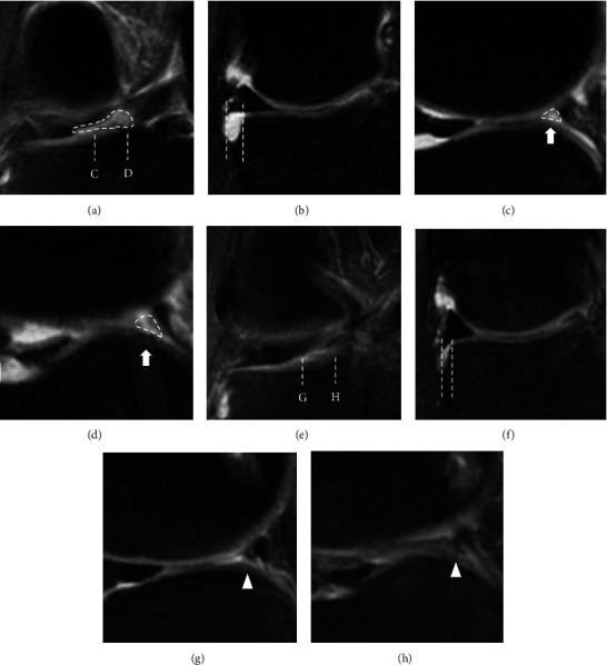
Lateral meniscus (LM) posterior root tear (LMPRT) is mainly caused by trauma, especially trauma associated with anterior cruciate ligament (ACL) injuries. Although a transtibial pullout repair or a side-to-side repair is commonly performed for LMPRT, to the best of our knowledge, there is no clinical report of LMPRT with tissue loss using the pullout technique. Thus, the purpose of this report was to describe a clinical, radiographic, and arthroscopic outcome after pullout repair for a case of LMPRT with a large defect with a chronic ACL tear and complex medial meniscus (MM) tears. A 31-year-old man complained of knee pain and restricted range of motion after twisting his knee when he stepped on an iron pipe. The patient had a football-related injury to his right knee 14 years before presentation, and since then, the patient's knee has given out more than 10 times but was left unassessed. Magnetic resonance imaging showed LMPRT with tissue loss, ACL tears, and complex MM tears. Transtibial pullout repair of the LMPRT with ACL reconstruction and MM repairs were performed. Following the pullout repair of the LMPRT, an approximately 6 mm gap remained between the LM posterior root and root insertion. However, magnetic resonance imaging and second-look arthroscopy at 1 year postoperatively revealed meniscal healing, gap filling with some regeneration tissue, of the LM posterior root. Furthermore, the lateral meniscus extrusion in the coronal plane improved from 3.1 mm (preoperative) to 1.6 mm (1 year postoperatively). Transtibial pullout repair with the remaining gap could be a viable treatment option for LMPRT with tissue loss, combined with ACL reconstruction.

 求助内容:
求助内容: 应助结果提醒方式:


