{"title":"A study of the chest imaging findings of adult patients with COVID-19 on admission to a tertiary hospital in Johannesburg, South Africa.","authors":"Ashleigh A Ord, Jarrod Zamparini, Liam Lorentz, Ashesh Ranchod, Halvani Moodley","doi":"10.4102/sajid.v37i1.449","DOIUrl":null,"url":null,"abstract":"<p><strong>Background: </strong>South Africa has experienced multiple waves of the coronavirus disease 2019 (COVID-19) with little research documenting chest imaging features in an human immunodeficiency virus (HIV) and tuberculosis (TB) endemic region.</p><p><strong>Objectives: </strong>Describe the chest imaging features, demographics and clinical characteristics of COVID-19 in an urban population.</p><p><strong>Method: </strong>Retrospective, cross-sectional, review of chest radiographs and computed tomographies (CTs) of adults admitted to a tertiary hospital with severe acute respiratory syndrome coronavirus 2 (SARS-CoV-2) infection, between 01 May 2020 and 30 June 2020. Imaging was reviewed by three radiologists. Clinical parameters and laboratory data were analysed.</p><p><strong>Results: </strong>A total of 113 adult patients with a mean age of 46 years and 10 months were included. A total of 113 chest radiographs and six CTs were read. Nineteen patients were HIV-positive (16.8%), 40 were hypertensive and diabetic (35.4%), respectively, and one had TB (0.9%). Common symptoms included cough (<i>n</i> = 69; 61.6%), dyspnoea (<i>n</i> = 60; 53.1%) and fever (<i>n</i> = 46; 40.7%). Lower zone predominant ground glass opacities (58.4%) and consolidation (29.2%) were most frequent on chest radiographs. The right lower lobe was most involved (46.9% ground glass opacities and 17.7% consolidation), with relative sparing of the left upper lobe. Bilateral ground glass opacities (66.7%) were most common on CT. Among the HIV-positive, ground glass opacities and consolidation were less common than in HIV-negative or unknown patients (<i>p</i> = 0.037 and <i>p</i> = 0.05, respectively).</p><p><strong>Conclusion: </strong>COVID-19 in South Africa has similar chest imaging findings to those documented globally, with some differences between HIV-positive and HIV-negative or unknown patients. The authors corroborate relative sparing of the left upper lobe; however, further research is required to validate this currently unique local finding.</p>","PeriodicalId":44007,"journal":{"name":"Southern African Journal of Infectious Diseases","volume":" ","pages":"449"},"PeriodicalIF":1.3000,"publicationDate":"2022-08-30","publicationTypes":"Journal Article","fieldsOfStudy":null,"isOpenAccess":false,"openAccessPdf":"https://www.ncbi.nlm.nih.gov/pmc/articles/PMC9452920/pdf/","citationCount":"0","resultStr":null,"platform":"Semanticscholar","paperid":null,"PeriodicalName":"Southern African Journal of Infectious Diseases","FirstCategoryId":"1085","ListUrlMain":"https://doi.org/10.4102/sajid.v37i1.449","RegionNum":0,"RegionCategory":null,"ArticlePicture":[],"TitleCN":null,"AbstractTextCN":null,"PMCID":null,"EPubDate":"2022/1/1 0:00:00","PubModel":"eCollection","JCR":"Q4","JCRName":"INFECTIOUS DISEASES","Score":null,"Total":0}
引用次数: 0
Abstract
Background: South Africa has experienced multiple waves of the coronavirus disease 2019 (COVID-19) with little research documenting chest imaging features in an human immunodeficiency virus (HIV) and tuberculosis (TB) endemic region.
Objectives: Describe the chest imaging features, demographics and clinical characteristics of COVID-19 in an urban population.
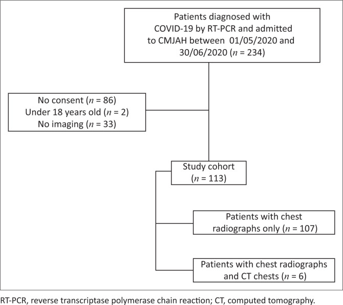
Method: Retrospective, cross-sectional, review of chest radiographs and computed tomographies (CTs) of adults admitted to a tertiary hospital with severe acute respiratory syndrome coronavirus 2 (SARS-CoV-2) infection, between 01 May 2020 and 30 June 2020. Imaging was reviewed by three radiologists. Clinical parameters and laboratory data were analysed.
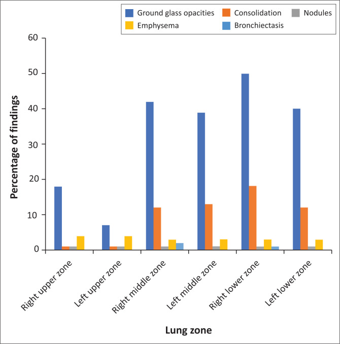
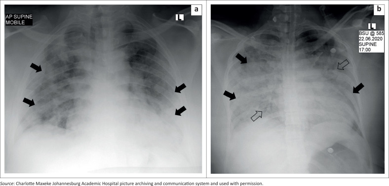
Results: A total of 113 adult patients with a mean age of 46 years and 10 months were included. A total of 113 chest radiographs and six CTs were read. Nineteen patients were HIV-positive (16.8%), 40 were hypertensive and diabetic (35.4%), respectively, and one had TB (0.9%). Common symptoms included cough (n = 69; 61.6%), dyspnoea (n = 60; 53.1%) and fever (n = 46; 40.7%). Lower zone predominant ground glass opacities (58.4%) and consolidation (29.2%) were most frequent on chest radiographs. The right lower lobe was most involved (46.9% ground glass opacities and 17.7% consolidation), with relative sparing of the left upper lobe. Bilateral ground glass opacities (66.7%) were most common on CT. Among the HIV-positive, ground glass opacities and consolidation were less common than in HIV-negative or unknown patients (p = 0.037 and p = 0.05, respectively).
Conclusion: COVID-19 in South Africa has similar chest imaging findings to those documented globally, with some differences between HIV-positive and HIV-negative or unknown patients. The authors corroborate relative sparing of the left upper lobe; however, further research is required to validate this currently unique local finding.
 求助内容:
求助内容: 应助结果提醒方式:


