{"title":"Diquafosol ophthalmic solution enhances mucin expression via ERK activation in human conjunctival epithelial cells with hyperosmotic stress.","authors":"Hyun Jung Lee, Soonwon Yang, Eun Jeong Cheon, Soojung Shin, Yong-Soo Byun, Hyun Seung Kim, So-Hyang Chung","doi":"","DOIUrl":null,"url":null,"abstract":"<p><strong>Purpose: </strong>To evaluate the effect of diquafosol tetrasodium on the expression of secretory and membrane-associated mucins in multi-layered cultures of primary human conjunctival epithelial cells (HCEC) using intracellular extracellular signal regulated kinase (ERK) signaling.</p><p><strong>Methods: </strong>HCECs were treated with hyperosmotic stress (400 mOsm/l) for 24 h after air-liquid interface cell culture followed by treatment with diquafosol. HCECs were stimulated for 1 h with or without PD98059, an ERK inhibitor, then treated with diquafosol for 6 h and 24 h. Mucin 1 (MUC1), mucin 16 (MUC16), and MUC5AC mRNA and protein expression levels were analyzed, and cell viability was detected using an MTT assay. Western blot analysis was used to examine p44/42 MAPK (Erk1/2) and phosphorylated p44/42 MAPK (Erk1/2) expression.</p><p><strong>Results: </strong>Hyperosmotic stressed HCECs demonstrated increased MUC5AC secretion and gene expression when treated with diquafosol. MUC1 mRNA levels increased significantly at 24 h (p<0.01), and expression of MUC16 mRNA levels increased at 6 h and were maintained until 24 h (p<0.05).There was no significant difference in cell viability compared to the control group. Immunostaining results for MUC1, MUC16, and MUC5AC in diquafosol tetrasodium-treated HCECs at 24 h showed more positive cells than in the control group. Phosphorylation of p44/42 MAPK (Erk1/2) signaling molecules significantly increased from 5 min to 60 min (p<0.05). The effects of diquafosol on mucin expressions in hyperosmotic stressed HCECs were significantly inhibited by PD98059, an ERK inhibitor, at 6 h and 24 h.</p><p><strong>Conclusions: </strong>ERK signaling may regulate the expression levels of MUC1, MUC16, and MUC5AC induced by diquafosol in hyperosmotic stressed HCECs.</p>","PeriodicalId":18866,"journal":{"name":"Molecular Vision","volume":" ","pages":"114-123"},"PeriodicalIF":1.4000,"publicationDate":"2022-06-30","publicationTypes":"Journal Article","fieldsOfStudy":null,"isOpenAccess":false,"openAccessPdf":"https://ftp.ncbi.nlm.nih.gov/pub/pmc/oa_pdf/40/bf/mv-v28-114.PMC9352363.pdf","citationCount":"0","resultStr":null,"platform":"Semanticscholar","paperid":null,"PeriodicalName":"Molecular Vision","FirstCategoryId":"3","ListUrlMain":"","RegionNum":3,"RegionCategory":"医学","ArticlePicture":[],"TitleCN":null,"AbstractTextCN":null,"PMCID":null,"EPubDate":"2022/1/1 0:00:00","PubModel":"eCollection","JCR":"Q4","JCRName":"BIOCHEMISTRY & MOLECULAR BIOLOGY","Score":null,"Total":0}
引用次数: 0
Abstract
Purpose: To evaluate the effect of diquafosol tetrasodium on the expression of secretory and membrane-associated mucins in multi-layered cultures of primary human conjunctival epithelial cells (HCEC) using intracellular extracellular signal regulated kinase (ERK) signaling.
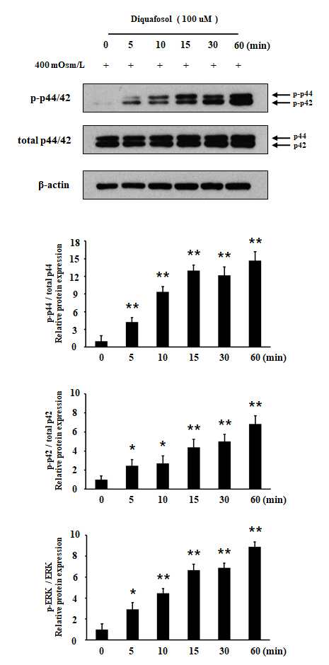
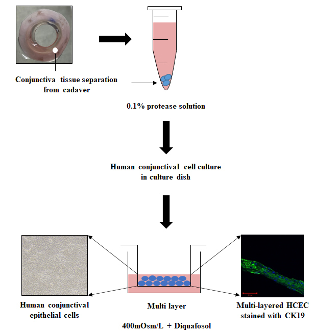
Methods: HCECs were treated with hyperosmotic stress (400 mOsm/l) for 24 h after air-liquid interface cell culture followed by treatment with diquafosol. HCECs were stimulated for 1 h with or without PD98059, an ERK inhibitor, then treated with diquafosol for 6 h and 24 h. Mucin 1 (MUC1), mucin 16 (MUC16), and MUC5AC mRNA and protein expression levels were analyzed, and cell viability was detected using an MTT assay. Western blot analysis was used to examine p44/42 MAPK (Erk1/2) and phosphorylated p44/42 MAPK (Erk1/2) expression.
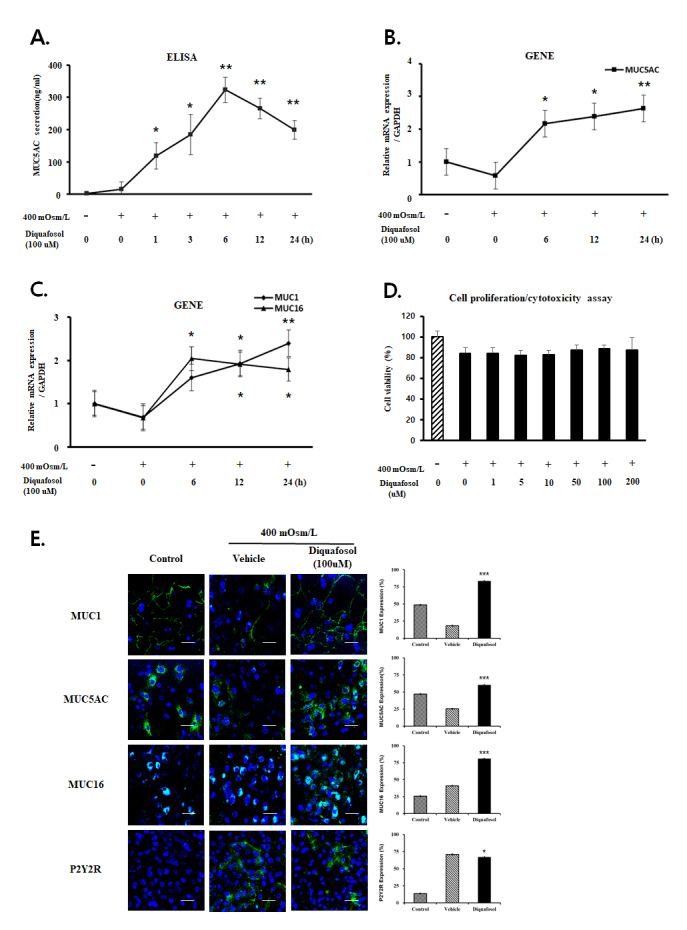
Results: Hyperosmotic stressed HCECs demonstrated increased MUC5AC secretion and gene expression when treated with diquafosol. MUC1 mRNA levels increased significantly at 24 h (p<0.01), and expression of MUC16 mRNA levels increased at 6 h and were maintained until 24 h (p<0.05).There was no significant difference in cell viability compared to the control group. Immunostaining results for MUC1, MUC16, and MUC5AC in diquafosol tetrasodium-treated HCECs at 24 h showed more positive cells than in the control group. Phosphorylation of p44/42 MAPK (Erk1/2) signaling molecules significantly increased from 5 min to 60 min (p<0.05). The effects of diquafosol on mucin expressions in hyperosmotic stressed HCECs were significantly inhibited by PD98059, an ERK inhibitor, at 6 h and 24 h.
Conclusions: ERK signaling may regulate the expression levels of MUC1, MUC16, and MUC5AC induced by diquafosol in hyperosmotic stressed HCECs.
期刊介绍:
Molecular Vision is a peer-reviewed journal dedicated to the dissemination of research results in molecular biology, cell biology, and the genetics of the visual system (ocular and cortical).
Molecular Vision publishes articles presenting original research that has not previously been published and comprehensive articles reviewing the current status of a particular field or topic. Submissions to Molecular Vision are subjected to rigorous peer review. Molecular Vision does NOT publish preprints.
For authors, Molecular Vision provides a rapid means of communicating important results. Access to Molecular Vision is free and unrestricted, allowing the widest possible audience for your article. Digital publishing allows you to use color images freely (and without fees). Additionally, you may publish animations, sounds, or other supplementary information that clarifies or supports your article. Each of the authors of an article may also list an electronic mail address (which will be updated upon request) to give interested readers easy access to authors.
 求助内容:
求助内容: 应助结果提醒方式:


