{"title":"Oligometastatic Squamous Cell Transformation From Metastatic Prostate Adenocarcinoma Treated With Systemic and Focal Therapy: A Case Report.","authors":"Karen Autio, Sean McBride","doi":"10.36401/JIPO-22-4","DOIUrl":null,"url":null,"abstract":"<p><p>Transformation to squamous cell carcinoma (SCC) after initial treatment of a primary prostate adenocarcinoma is rare and typically results in rapid treatment-refractory disease progression and death. Here, we present a case of a 70-year-old man who was initially treated with prostatectomy and radiotherapy, and later developed bone metastases. After commencing systemic therapy with androgen deprivation therapy (ADT) and apalutamide, his prostate-specific antigen (PSA) declined to undetectable levels, yet short-interval imaging demonstrated oligo-progression at T4, with biopsy specimen demonstrating pure SCC. Molecular profiling of both the primary prostate tumor and T4 demonstrated alterations in <i>TMPRSS2-ERG</i>, <i>TP53</i>, and <i>FOXA1</i> confirming site of origin, with loss of <i>RNF43</i> in the squamous metastasis. He was treated with stereotactic body radiation therapy to the SCC metastasis and continued on ADT and apalutamide with stable disease for a year post-radiation. This case highlights the importance of imaging to detect non-PSA-producing metastatic disease, the utility of radiation therapy in oligo-progression, and use of molecular profiling to provide insights into the pathogenesis of histologic transformation.</p>","PeriodicalId":16081,"journal":{"name":"Journal of Immunotherapy and Precision Oncology","volume":" ","pages":"79-83"},"PeriodicalIF":3.2000,"publicationDate":"2022-06-15","publicationTypes":"Journal Article","fieldsOfStudy":null,"isOpenAccess":false,"openAccessPdf":"https://ftp.ncbi.nlm.nih.gov/pub/pmc/oa_pdf/3b/7e/i2590-017X-5-3-79.PMC9390704.pdf","citationCount":"0","resultStr":null,"platform":"Semanticscholar","paperid":null,"PeriodicalName":"Journal of Immunotherapy and Precision Oncology","FirstCategoryId":"1085","ListUrlMain":"https://doi.org/10.36401/JIPO-22-4","RegionNum":0,"RegionCategory":null,"ArticlePicture":[],"TitleCN":null,"AbstractTextCN":null,"PMCID":null,"EPubDate":"2022/8/1 0:00:00","PubModel":"eCollection","JCR":"Q3","JCRName":"Medicine","Score":null,"Total":0}
引用次数: 0
Abstract
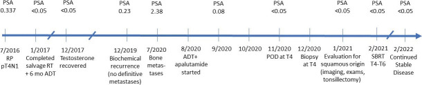
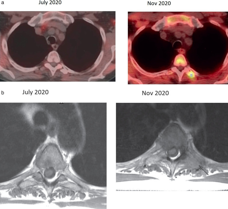
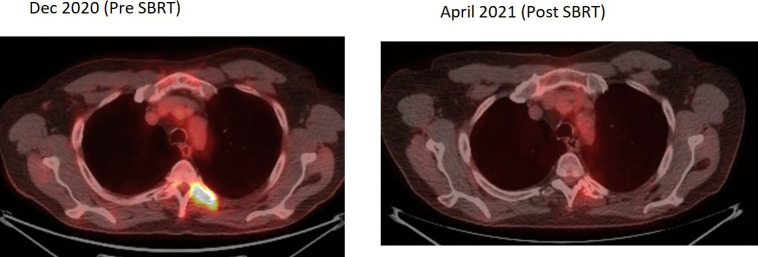
Transformation to squamous cell carcinoma (SCC) after initial treatment of a primary prostate adenocarcinoma is rare and typically results in rapid treatment-refractory disease progression and death. Here, we present a case of a 70-year-old man who was initially treated with prostatectomy and radiotherapy, and later developed bone metastases. After commencing systemic therapy with androgen deprivation therapy (ADT) and apalutamide, his prostate-specific antigen (PSA) declined to undetectable levels, yet short-interval imaging demonstrated oligo-progression at T4, with biopsy specimen demonstrating pure SCC. Molecular profiling of both the primary prostate tumor and T4 demonstrated alterations in TMPRSS2-ERG, TP53, and FOXA1 confirming site of origin, with loss of RNF43 in the squamous metastasis. He was treated with stereotactic body radiation therapy to the SCC metastasis and continued on ADT and apalutamide with stable disease for a year post-radiation. This case highlights the importance of imaging to detect non-PSA-producing metastatic disease, the utility of radiation therapy in oligo-progression, and use of molecular profiling to provide insights into the pathogenesis of histologic transformation.

 求助内容:
求助内容: 应助结果提醒方式:


