A systematic examination of brain volumetric abnormalities in recent-onset schizophrenia using voxel-based, surface-based and region-of-interest-based morphometric analyses.
John P John, Ammu Lukose, Bhavani Shankara Bagepally, Harsha N Halahalli, Nagaraj S Moily, Anupa A Vijayakumari, Sanjeev Jain
{"title":"A systematic examination of brain volumetric abnormalities in recent-onset schizophrenia using voxel-based, surface-based and region-of-interest-based morphometric analyses.","authors":"John P John, Ammu Lukose, Bhavani Shankara Bagepally, Harsha N Halahalli, Nagaraj S Moily, Anupa A Vijayakumari, Sanjeev Jain","doi":"10.1186/s12952-015-0030-z","DOIUrl":null,"url":null,"abstract":"<p><strong>Background: </strong>Brain morphometric abnormalities in schizophrenia have been extensively reported in the literature. Whole-brain volumetric reductions are almost universally reported by most studies irrespective of the characteristics of the samples studied (e.g., chronic/recent-onset; medicated/neuroleptic-naïve etc.). However, the same cannot be said of the reported regional morphometric abnormalities in schizophrenia. While certain regional morphometric abnormalities are more frequently reported than others, there are no such abnormalities that are universally reported across studies. Variability of socio-demographic and clinical characteristics across study samples as well as technical and methodological issues related to acquisition and analyses of brain structural images may contribute to inconsistency of brain morphometric findings in schizophrenia. The objective of the present study therefore was to systematically examine brain morphometry in patients with recent-onset schizophrenia to find out if there are significant whole-brain or regional volumetric differences detectable at the appropriate significance threshold, after attempting to control for various confounding factors that could impact brain volumes.</p><p><strong>Methods: </strong>Structural magnetic resonance images of 90 subjects (schizophrenia = 45; healthy subjects = 45) were acquired using a 3 Tesla magnet. Morphometric analyses were carried out following standard analyses pipelines of three most commonly used strategies, viz., whole-brain voxel-based morphometry, whole-brain surface-based morphometry, and between-group comparisons of regional volumes generated by automated segmentation and parcellation.</p><p><strong>Results: </strong>In our sample of patients having recent-onset schizophrenia with limited neuroleptic exposure, there were no significant whole brain or regional brain morphometric abnormalities noted at the appropriate statistical significance thresholds with or without including age, gender and intracranial volume or total brain volume in the statistical analyses.</p><p><strong>Conclusions: </strong>In the background of the conflicting findings in the literature, our findings indicate that brain morphometric abnormalities may not be directly related to the schizophrenia phenotype. Analysis of the reasons for the inconsistent results across studies as well as consideration of alternate sources of variability of brain morphology in schizophrenia such as epistatic and epigenetic mechanisms could perhaps advance our understanding of structural brain alterations in schizophrenia.</p>","PeriodicalId":73849,"journal":{"name":"Journal of negative results in biomedicine","volume":"14 ","pages":"11"},"PeriodicalIF":0.0000,"publicationDate":"2015-06-12","publicationTypes":"Journal Article","fieldsOfStudy":null,"isOpenAccess":false,"openAccessPdf":"https://sci-hub-pdf.com/10.1186/s12952-015-0030-z","citationCount":"9","resultStr":null,"platform":"Semanticscholar","paperid":null,"PeriodicalName":"Journal of negative results in biomedicine","FirstCategoryId":"1085","ListUrlMain":"https://doi.org/10.1186/s12952-015-0030-z","RegionNum":0,"RegionCategory":null,"ArticlePicture":[],"TitleCN":null,"AbstractTextCN":null,"PMCID":null,"EPubDate":"","PubModel":"","JCR":"","JCRName":"","Score":null,"Total":0}
引用次数: 9
Abstract
Background: Brain morphometric abnormalities in schizophrenia have been extensively reported in the literature. Whole-brain volumetric reductions are almost universally reported by most studies irrespective of the characteristics of the samples studied (e.g., chronic/recent-onset; medicated/neuroleptic-naïve etc.). However, the same cannot be said of the reported regional morphometric abnormalities in schizophrenia. While certain regional morphometric abnormalities are more frequently reported than others, there are no such abnormalities that are universally reported across studies. Variability of socio-demographic and clinical characteristics across study samples as well as technical and methodological issues related to acquisition and analyses of brain structural images may contribute to inconsistency of brain morphometric findings in schizophrenia. The objective of the present study therefore was to systematically examine brain morphometry in patients with recent-onset schizophrenia to find out if there are significant whole-brain or regional volumetric differences detectable at the appropriate significance threshold, after attempting to control for various confounding factors that could impact brain volumes.
Methods: Structural magnetic resonance images of 90 subjects (schizophrenia = 45; healthy subjects = 45) were acquired using a 3 Tesla magnet. Morphometric analyses were carried out following standard analyses pipelines of three most commonly used strategies, viz., whole-brain voxel-based morphometry, whole-brain surface-based morphometry, and between-group comparisons of regional volumes generated by automated segmentation and parcellation.
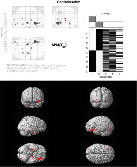
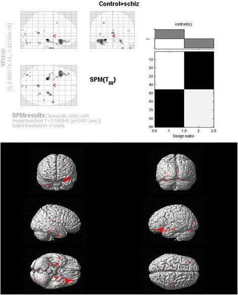
Results: In our sample of patients having recent-onset schizophrenia with limited neuroleptic exposure, there were no significant whole brain or regional brain morphometric abnormalities noted at the appropriate statistical significance thresholds with or without including age, gender and intracranial volume or total brain volume in the statistical analyses.
Conclusions: In the background of the conflicting findings in the literature, our findings indicate that brain morphometric abnormalities may not be directly related to the schizophrenia phenotype. Analysis of the reasons for the inconsistent results across studies as well as consideration of alternate sources of variability of brain morphology in schizophrenia such as epistatic and epigenetic mechanisms could perhaps advance our understanding of structural brain alterations in schizophrenia.


 求助内容:
求助内容: 应助结果提醒方式:


